 1300 164 990
Fluorescein Angiography: Side Effects, Procedure, Risk and Benefits
Published on May 29th, 2024
 Australia

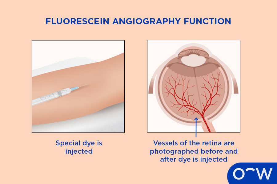
A fluorescein angiography is a test that involves capturing images of the retina, which is located in the back of the eye. During a fluorescein angiography, the ophthalmologist will need to dilate your pupils to widen them and inject a dye known as fluorescein through your arm. The fluorescent dye then travels to the blood vessels in the eye and highlights your blood vessels, helping the optometrist to examine them and check for any signs of problems that may impair your vision. A fluorescein dye test is helpful for screening eye conditions such as age-related macula degeneration, cystoid macula edema, diabetes-related retinopathy, macula hole, macula pucker, ocular melanoma, retinal detachment and retinitis pigmentosa.
What is Fluorescein Angiography?
A fluorescein angiography is a type of test that involves a special dye and camera to examine the back region of the eye known as the retina. A fluorescein angiography will first involve taking one round of images before injecting the fluorescein dye into your arm. This will then enable the dye to travel to the blood vessels in the eye making them easier to examine. The optometrist will then take another round of photos of the retina once they have been highlighted. A fluorescein dye test is normally performed by an ophthalmologist after having an eye examination or eye test with an optometrist first.
How Does a Fluorescein Angiography Test Work?
A fluorescein angiography test works by using a special contrast dye called fluorescein to highlight the blood vessels in your eye. A fluorescein angiography helps the ophthalmologist assess the physical condition and circulation of the retina and choroid according to Ruia S. and Koushik T. (2023).
What are the Fluorescein Angiography Side Effects?
The fluorescein angiography side effects may include blurry vision and increased light sensitivity. Blurry vision and light sensitivity can ensue after having a fluorescein angiography as a result of the dilating eye drops that is used and may last from 12 to 24 hours.
How is Fluorescein Angiography Done?
A fluorescein angiography is done by first injecting a contrast fluorescein dye in the vein of your arm. The dye then passes through your bloodstream reaching the blood vessels in your eyes after a short period. The ophthalmologist will then capture images of your eyes while the dye is in the retina and will assist with revealing any abnormalities in your blood vessels. A fluorescein angiography procedure may take around 20 minutes and is generally performed if your eye condition indicates signs of a retinal disease.
What Should the Eye Doctor Consider Before Conducting a Fluorescein Angiography?
The eye doctor should consider your medical history and recent eye test results before conducting a fluorescein angiography. The eye doctor should consider your medical history to ensure they are informed of any possible allergies you may have and to indicate if there has been a history of retinal diseases in your family. The results of your recent eye test should also be taken into consideration as it will indicate the relevant signs of an eye problem. The eye care specialist that will conduct this test will be an ophthalmologist. An ophthalmologist is a medical doctor with a specialisation in eye care. Ophthalmologists are trained to diagnose and treat eye conditions as well as perform eye surgery.
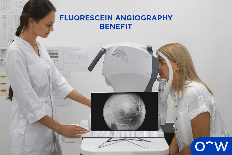
What are the Benefits of Fluorescein Angiography?
The benefit of a fluorescein angiography is its assistance with evaluating the condition of the blood vessels in your eye and the circulation of your blood flow in the retina and choroid. A fluorescein angiography is necessary for identifying abnormal blood vessel growth and other retinal problems according to the American Academy of Ophthalmology.
What is the Risk of Fluorescein Angiography?
The risk of fluorescein angiography is having an allergic reaction to the dye. The risk of having an allergic reaction to the fluorescein dye is considered low risk according to the Cleveland Clinic. Individuals who may be allergic to the dye, may experience hives, itchy skin or difficulty breathing according to the American Academy of Ophthalmology.
What Can Fluorescein Angiography Detect?
A fluorescein angiography can detect age-related macula degeneration, cystoid macula edema, diabetes-related retinopathy, macula hole, macula pucker, ocular melanoma, retinal detachment and retinitis pigmentosa. The eye conditions that a fluorescein angiography can detect are listed below.
- Age-related Macular Degeneration: Age-related macula degeneration refers to an eye disease in which damage to cells in the macula lead to loss of central vision. Symptoms of age-related macula degeneration may include blurry vision, straight lines appearing wavy, dark spots appearing in central vision and loss of central vision according to John Hopkins Medicine. Treating wet age-related macula degeneration may require anti-VEGF injections according to the National Eye Institute. Age-related macula degeneration can develop as a result of natural changes to structures and blood circulation as you age.
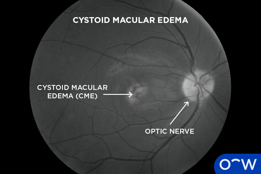
- Cystoid Macular Edema: Cystoid macula edema is a painless condition that affects the macula. Cystoid Macula edema can stem from causes such as inflammation from eye surgery, diabetes, blockage in your retinal veins, eye injury and side effects of certain medications. Symptoms of cystoid macula edema include blurry or distorted central vision, different appearance of colours and the dim or dark appearance of objects in view.
- Diabetes-Related Retinopathy: Diabetic retinopathy is a complication of diabetes that affects the blood vessels in the retina. Damage to these blood vessels can lead to vision loss and can eventually lead to blindness if untreated. There are no symptoms in early-stage diabetic retinopathy but later-stage symptoms include fluctuating vision, increased light sensitivity, glare and eye floaters. Treatments for diabetic retinopathy include medication, laser treatment and eye surgery.
- Macula Hole: A macula hole refers to a hole, tear or break in the macula, which is located in the retina of the eye. A hole in this part of the retina is commonly caused by vitreous detachment, in which the vitreous fluid does not fully separate from the macula, leaving a hole or tear, and is often caused by ageing. A macula hole may be treated with a surgical process known as a vitrectomy.
- Macular Pucker: Macular pucker also known as an epiretinal membrane is a rare eye condition that can cause your vision to appear wavy or distorted and occurs when wrinkles or creases develop on the macula. The macula should be in a flat position at the back of your eye to function correctly, therefore, wrinkles and creases forming can impair your vision. Symptoms may include difficulty seeing details, and a grey or cloudy area in your central vision. Treating macula pucker can include a vitrectomy for severe symptoms. For mild symptoms you may not require treatment or you may just need to have your prescription updated as eye drops and laser surgery will not be effective.
- Ocular Melanoma: Ocular Melanoma or eye melanoma, is a form of cancer that occurs in the eye. Melanoma is a type of cancer that affects melanin-producing cells, which are responsible for giving the body its skin colour. The causes of ocular melanoma are not definitively known, however, there are several risk factors of ocular melanoma which can include having a fair complexion, having light-coloured eyes, being of older age, abnormal growths or lesions on the eye, a family or personal history of melanoma and certain skin conditions.
- Retinal Detachment: Retinal detachment is considered a medical emergency. That happens when the retina pulls away from its normal position at the back of your eye. Symptoms of retinal detachment are an increase of floaters, light flashes in one or both eyes and a dark curtain-like shadow on the sides of your vision. You may be more at risk of developing retinal detachment if there is a history of retinal detachment in your family or if you have had a severe eye injury or surgery.
- Retinitis Pigmentosa: Retinitis pigmentosa refers to a group of rare eye diseases that cause damage to the retina potentially caused by genetics. The retina is the tissue layer of photoreceptor cells and glial cells located at the back of the eye and is responsible for processing the light that enters the eye and converts it to the images you see. Symptoms of retinitis pigmentosa can include low light vision problems, poor night vision and glare sensitivity.
What are the Stages of Fluorescein Angiography?
The stages of fluorescein angiography include the choroidal phase, arterial phase, arteriovenous, venous and recirculation. The stages of fluorescein angiography are listed below.
- Choroidal Phase: The choroidal phase is where the dye takes under 20 seconds to reach the retina. The posterior ciliary arteries is the first region that the dye fills, therefore establishing a choroidal filling according to Ruia, S. and Tripathy, K. (2023).
- Arterial Phase: The arterial phase refers to when the dye moves to the arteries which can be as quick as 1 or 2 seconds after the choroidal phase.
- Venous: Venous involves three phases, early, mid and late depending on the filling of the fluorescein dye. The early phase exhibits a smooth flow, and then complete fulfilment of the dye in the veins in the mid phase, which is then followed by less dye in the arteries during the late phase.
- Recirculation: Recirculation is the occurrence after the venous phase, wherein, the dye begins reducing in the vessels and clearing out at around 10 minutes according to the National Library of Medicine.
Is a Fluorescein Angiography Necessary?
Yes, a fluorescein angiography is necessary if your eye test indicates signs of retinal problems or abnormal vessel growth. It may not be necessary for patients who do not exhibit any problems in their eye condition, however, your optometrist will confirm your results when you first get an eye test.
Does a Fluorescein Angiography Cause Temporary Yellow Vision?
No, fluorescein angiography won’t cause temporary yellow vision. Fluorescein angiography may instead lead to a yellowish-tinge on skin from the dye injection. The dilating eye drops may lead to blurry vision and light sensitivity for 12-24 hours.
Read Fluorescein Angiography: Side Effects, Procedure, Risk and Benefits in other Oscar Wylee regions and their languages.
Australia