


 1300 164 990
Vitreomacular Traction (VMT): Symptoms and Causes
Published on October 8th, 2026
Vitreomacular traction (VMT) is a potentially sight-threatening condition where the vitreous gel fails to cleanly detach from the macula and instead pulls on it. VMT symptoms include blurred vision, distorted vision, micropsia, scotoma, and reading difficulty. Aging is the primary risk factor for VMT, as the vitreous gel naturally shrinks and liquefies over time. Treatments for VMT range from close monitoring to injections and surgery in severe cases.
It is important to note that Oscar Wylee offers optometry services only; this article is for educational purposes only and is not a substitute for professional medical advice, diagnosis, or treatment.
What is Vitreomacular Traction?
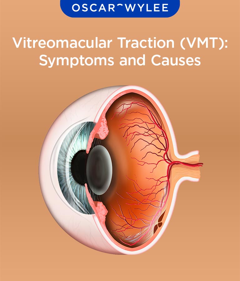
Vitreomacular Traction (VMT) occurs when the vitreous gel (jelly-like substance) fails to cleanly detach from the macula and instead pulls and tugs on this central part of the retina, according to the Macular Disease Foundation Australia. The macula is a sensitive area at the centre of the retina responsible for sharp central vision. When the macula is pulled by the vitreous, it distorts the macula's shape and can cause vision changes.
How does Vitreomacular Traction Affect the Eye?
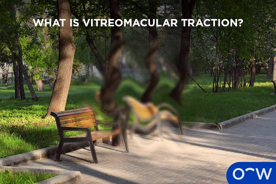
VMT causes the vitreous to tug on the macula, which distorts its shape, causes swelling, and impairs vision, according to the Macular Disease Foundation Australia. This pulling stretches and splits the retinal layers, causing fluid collection and swelling. Scar tissue can form on the macula, which leads to wrinkling of the retinal tissue. If tugging is severe, it can cause a hole in the centre of the retina, called a macular hole, which significantly reduces vision.
Is VMT Caused by Vitreous Pulling on the Macula?
Yes, VMT is directly caused by the vitreous pulling on the macula, according to the Macular Disease Foundation Australia.
What Causes Vitreomacular Traction in the Eye?
VMT is caused by incomplete posterior vitreous detachment (PVD), as the eye ages. As people age, the vitreous gel shrinks and becomes more watery. In normal vitreous separation, the vitreous gel pulls away from the retina, however, in VMT, the gel fails to separate cleanly and remains stuck to the macula, and causes a tugging force on the macula which distorts its shape, according to the Macular Disease Foundation Australia. Apart from old age, those at higher risk of VMT are people with high myopia, age-related macular degeneration (AMD), diabetic eye disease, retinal vein occlusion, and ocular trauma.
Which Eye Changes Can Lead to VMT?
Eye changes that can lead to VMT are incomplete posterior vitreous detachment (PVD), age-related vitreous changes, structural and mechanical preconditions, and underlying retinal and vascular diseases. These eye changes that can lead to VMT are explained below.
- Incomplete Posterior Vitreous Detachment (PVD): Incomplete posterior vitreous detachment (PVD) is the primary cause of VMT. Incomplete PVD occurs when the gel-like vitreous in the middle of the eye separates mostly from the retina, but remains abnormally stuck to the macula, according to the MDFA.
- Age-Related Vitreous Changes: Age-related vitreous changes refers to the vitreous gel becoming more watery and fibrous as you age, causing it to collapse forward. This can increase the risk of VMT.
- Structural and Mechanical Preconditions: Structural and mechanical preconditions refer to high myopia, epiretinal membrane (ERM) formation, and prior retinal surgery or ocular trauma.
- Underlying Retinal and Vascular Diseases: Underlying retinal and vascular diseases such as diabetic retinopathy, age-related macular degeneration (AMD), and retinal vein occlusion (RVO) can increase the risk of developing VMT.
Can Posterior Vitreous Detachment Cause VMT?
Yes, an incomplete posterior vitreous detachment (PVD) is the primary cause of vitreomacular traction (VMT), according to the Macular Disease Foundation Australia (MDFA).
What are the Common Vitreomacular Traction Symptoms?
The common VMT symptoms are blurred vision, distorted vision, micropsia, scotoma, and photopsia. Common VMT symptoms are explained below.
- Blurred Vision: Blurred vision refers to a gradual or sudden decrease in vision, making daily tasks difficult.
- Distorted Vision: Distorted vision involves straight lines appearing abnormally bent or wavy, according to Perth Retina.
- Micropsia: Micropsia is when objects appear smaller than they are.
- Scotoma: Scotoma refers to blind spots in the field of vision.
- Photopsia: Photopsia is brief flashes of light in the field of vision.
1. Blurred Vision
Blurred vision can be caused by VMT because the eye's vitreous gel abnormally sticks to and physically pulls on the macula, warping its structure and disrupting its ability to process light, according to the MDFA. Blurred vision makes daily tasks, such as reading, difficult.
2. Distorted Vision
Distorted vision makes straight lines appear bent, crooked, or wavy. VMT causes distorted vision in the same way it causes blurred vision, as the warped structure of the macula disrupts normal light processing.
3. Micropsia
Micropsia is when objects appear smaller than they are. VMT causes micropsia because the mechanical tugging on the macula stretches and spreads the retinal photoreceptors further apart, according to the Royal Australian College of General Practitioners (RACGP).
4. Scotoma
Scotoma is a central blind spot in the field of vision. VMT causes scotoma due to the physical pulling forces that structurally affect the macula.
5. Photopsia
Photopsia refers to brief flashes of light in the field of vision. VMT causes photopsia due to the mechanical pulling of the vitreous gel on the retina, according to the MDFA.
How Is Vitreomacular Traction Diagnosed?
VMT is primarily diagnosed by an optometrist or ophthalmologist through a comprehensive eye test and confirmed through an OCT scan, according to the MDFA. VMT is difficult to diagnose through a physical examination alone, and an OCT scan is the gold standard test to diagnose VMT. An OCT scan is a quick, non-invasive scan that uses light waves to capture ultra-high-resolution, cross-sectional images of the retina's distinct tissue layers, according to the MDFA. If an optometrist detects VMT on an OCT scan, they will refer you to an ophthalmologist (retinal specialist) for further medical evaluation, diagnosis, and specialised care. A dilated retina exam (funduscopy) can also be performed by an optometrist to examine the retina, macula, and vitreous gel in detail.
What does VMT Look Like on an OCT Scan?
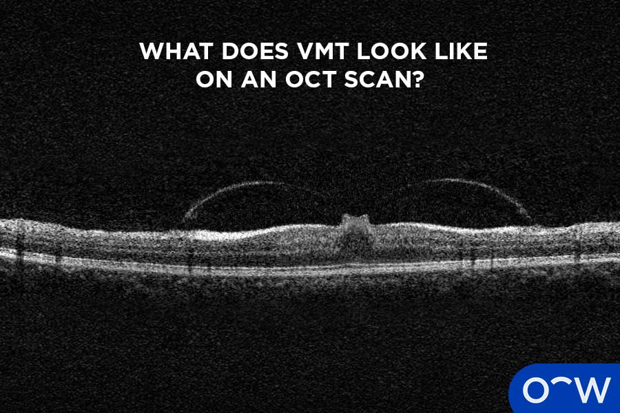
On an OCT scan, VMT looks like an incomplete or partial separation of the vitreous gel from the retina, causing a clear, visible, physical pulling on the retina. Incomplete PVD, persistent central attachment, and retinal distortion will all be visible to the optometrist or ophthalmologist performing the scan.
Can OCT Confirm Vitreomacular Traction?
Yes, OCT is the primary imaging method used to confirm VMT. OCT is the gold standard test performed by an optometrist or ophthalmologist diagnosing VMT.
How is Vitreomacular Adhesion Different From VMT?
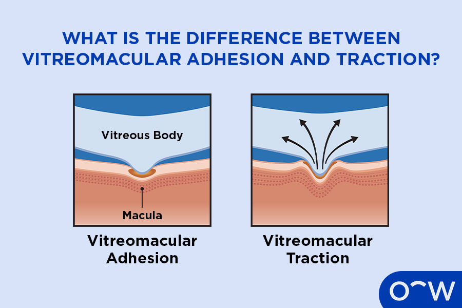
Vitreomacular adhesion (VMA) differentiates from VMT based on whether the attached eye gel is causing physical damage or distortion to the retina. Both conditions occur during the natural aging process when the eye's vitreous gel fails to separate cleanly from the macula, according to the MDFA. In VMA, the vitreous gel is attached to the macula but does not pull or distort it, and retinal layers look completely normal on an OCT scan. VMA also usually causes no symptoms, and it is considered a normal stage of eye aging, and not a disease. VMA can often resolve on its own without intervention, whereas VMT requires specialist monitoring and potentially, surgery.
Can Vitreomacular Adhesion Progress to VMT?
Yes, VMA can progress to VMT. However, Australian clinical data confirms that VMA is highly common, affecting roughly 15% to 30% of adults over the age of 45, whereas VMT is significantly rarer, affecting about 1% of the population.
Can Vitreomacular Traction Resolve on Its Own?
Yes, VMT can resolve on its own, but this only happens in a low to moderate number of cases. If adhesion is minor, there is no underlying scar tissue, and/or the eye still has its natural lens, resolution without intervention is more likely. If VMT involves broad adhesion, severe vision loss, or a macular hole, surgical intervention is required. However, if VMT is mild without major symptoms such as blurred vision, an optometrist or ophthalmologist may recommend just monitoring the condition with regular check-ups.
When Does VMT Need Treatment?
VMT needs treatment when symptoms appear, progressive vision loss occurs, or if it leads to structural retinal damage, according to the MDFA. While an optometrist can monitor mild cases during routine check-ups, an ophthalmologist will decide if active medical intervention is required. Treatment is recommended if there is a clear impact on daily activities, progressive vision loss, or secondary retinal complications, such as a macular hole. If not, VMT can be left without treatment with close monitoring by an optometrist or ophthalmologist.
What Treatments Are Used for Vitreomacular Traction?
The treatments used for VMT are observation, medication, pneumatic vitreolysis, and vitrectomy surgery. These treatments for VMT are explained below.
- Observation: Observation is used for mild or asymptomatic cases of VMT. Observation involves an optometrist or ophthalmologist regularly checking your eyes using an OCT.
- Medication: Medication for VMT refers to pharmacological vitreolysis (eye injections), which is a singular injection of a human enzyme by an ophthalmologist that dissolves the proteins stuck to the vitreous gel of the macula, according to the Australian Prescriber.
- Pneumatic Vitreolysis: Pneumatic vitreolysis refers to a small gas bubble being injected into the eye by an ophthalmologist and the patient performing specific exercises that can help break off the adhesion.
- Vitrectomy Surgery: Vitrectomy surgery involves an ophthalmic surgeon entering the eye to remove the tugging vitreous jelly, sometimes placing a temporary gas bubble inside the eye to help the retina settle.
When Is Surgery Considered for VMT?
Surgery is considered for VMT if it turns into a progressive, sight-threatening condition that affects daily life. When secondary retinal damage occurs, such as a macular hole or macular pucker, surgery is strongly recommended. If retinal splitting or thickening is apparent, then surgery is also preferred. Lastly, when daily activities such as reading, driving, and recognising faces are affected, surgery is also considered.