


 1300 164 990
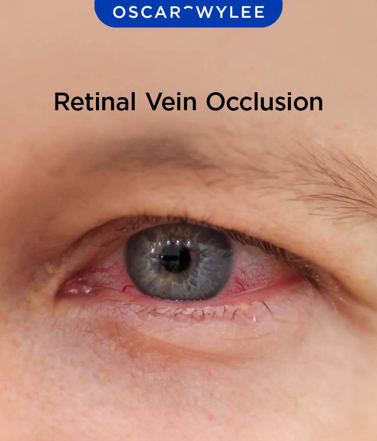
Retinal Vein Occlusion (RVO): Symptoms, Causes and Treatment
Published on February 24th, 2026
A retinal vein occlusion (RVO) occurs when one of the veins in your retina becomes blocked. As a result, a person can experience varying degrees of vision loss depending on the severity and location of the block, according to the Macular Disease Foundation Australia. Read on for more about Retinal Vein Occlusion (RVO), including its symptoms, causes and treatment options.
What Is Retinal Vein Occlusion (RVO)?
A retinal vein occlusion refers to a blockage of one of the veins that drains blood from the eye. This can lead to reduced blood flow, retinal swelling, retinal hemorrhages and reduced oxygen supply to retinal tissues.
How Does Retinal Vein Occlusion Affect Vision?
Retinal vein occlusion can affect vision as those affected may notice sudden or gradual painless loss of vision or visual field defects in their periphery or central vision, according to The College of Optometrists. The impact of vision will vary depending on the location and severity of the blockage. Some people experience noticeable symptoms, while others may have minimal changes.
Can Retinal Vein Occlusion Cause Permanent Vision Loss?
Yes, retinal vein occlusion in some cases can cause permanent vision loss or lasting visual changes in some individuals, depending on the severity of the vein blockage. Early treatment can help to reduce the risk of long term vision loss.
What Are the Symptoms of Retinal Vein Occlusion?
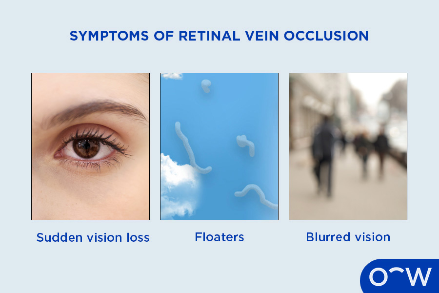
The symptoms of retinal vein occlusion include blurred vision, floaters in some cases, and gradual or sudden vision loss, according to the Eye Specialist Institute. Most of these symptoms may be painless and vary from person to person, if there are any unexplained changes to your vision, it is important to consult an optometrist.
How Can You Recognise a Blood Clot in the Eye in Retinal Vein Occlusion (RVO)?
You can recognise a blood clot in the eye in Retinal Vein Occlusion (RVO) by the effect it will have on your vision, as it can lead to varying degrees of vision loss. The presence of a blood clot will be best checked by an optometrist.
Does Retinal Vein Occlusion (RVO) Lead to Blurred Vision?
Yes, retinal vein occlusion can lead to blurred vision or complete vision loss. Whether you have blurred vision or complete blindness will depend on the blockage location and its severity.
What Causes Retinal Vein Occlusion (RVO)?
The cause of retinal vein occlusion is due to blood flow within a retinal vein becoming obstructed, primarily due to clot formation/vessel wall changes.
Which Medical Conditions Increase the Risk of Retinal Vein Occlusion (RVO)?
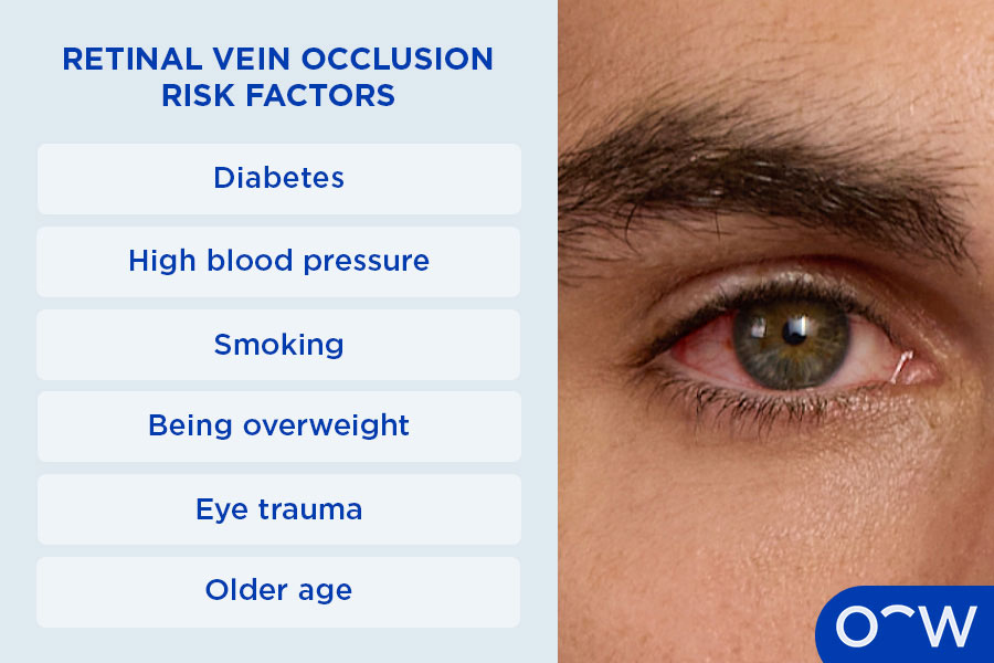
The medical conditions that can increase the risk of retinal vein occlusion (RVO) include hypertension, diabetes, glaucoma and age, according to Armadale Eye Clinic. It is important to manage these conditions by regularly checking in with your doctor and implementing their advice.
Can Retinal Vein Occlusion (RVO) Happen in Healthy Children or Adults?
It is rare for retinal vein occlusion (RVO) to happen in healthy children or adults. Children may experience it due to inflammatory or blood-clotting conditions. Typically, older adults with health conditions such as hypertension or glaucoma will be more at risk of getting retinal vein occlusion.
What Are the Different Types of Retinal Vein Occlusion (RVO)?
The different types of retinal vein occlusion (RVO) are central retinal vein occlusion and branch retinal vein occlusion. These two types affect the eye in different ways. Read more about the different types of retinal vein occlusion below.
- Central Retinal Vein Occlusion (CRVO): Central Retinal Vein Occlusion (CRVO) occurs when the main retinal vein becomes blocked.
- Branch Retinal Vein Occlusion (BRVO): Branch Retinal Vein Occlusion (BRVO) occurs due to the blockage of a smaller retinal branch.
Central Retinal Vein Occlusion
Central Retinal Vein Occlusion (CRVO) is the result of blood clots blocking the main vessel for draining blood from the retina. This affects larger portions of the retina, leading to more severe vision changes.
Branch Retinal Vein Occlusion (BRVO)
Branch Retinal Vein Occlusion (BRVO) is the blockage of blood flow in the area where the branch retinal vein is crossed by a branch retinal artery. This affects a smaller part of the retina.
Can Branch Retinal Vein Occlusion (BRVO) Lead to Vision Loss?
Yes, branch retinal vein occlusion can lead to vision loss that is often painless and unexpected.
How Is Retinal Vein Occlusion (RVO) Diagnosed?
Retinal vein occlusion (RVO) is diagnosed by an optometrist, and you will be referred to an ophthalmologist for treatment according to the Macular Disease Foundation Australia. The diagnosis process involves the standard eye test and specialised eye tests such as an Ophthalmoscopy, Optical Coherence Tomography (OCT), Fluorescein Angiography and blood tests according to Armadale Eye Clinic.
Which Eye Tests Can Detect Retinal Vein Occlusion (RVO)?
The eye tests that can detect Retinal Vein Occlusion (RVO) are a Dilated retinal examination (fundoscopy), Optical Coherence Tomography (OCT), Fluorescein Angiography in some cases, and blood tests. An Ophthalmoscopy test is when a bright light is shone toward the back of the eye to observe the retina and retinal blood vessels. An Optical Coherence Tomography provides cross-sectional images of the retina to show swelling or fluid that may be present. Fluorescein Angiography refers to a test done where fluorescent dye is put into the bloodstream to illuminate the identification of potential blockages within retinal vessels. Blood tests are conducted to identify a potential cause of the retinal vein occlusion, such as diabetes or blood-clotting disorders.
Is a Fundoscopy Required to Diagnose Retinal Vein Occlusion (RVO)?
Yes, a fundoscopy is often required to diagnose retinal vein occlusion (RVO). This process involves an eye specialist examining the back of the eye with a special magnifying lens to assess whether there is bleeding or foreign cells and fluids in the retina.
What Are the Treatment Options for Retinal Vein Occlusion (RVO)?
The treatment options for retinal vein occlusion (RVO) include Anti-VEGF Injections, vitrectomy surgery or steroid injections. Read more about these treatment options for retinal vein occlusion (RVO) below.
- Anti-VEGF Injections: Anti-VEGF injections are directly injected into the eyeball and prevent the growth of abnormal new blood vessels, often repeated monthly, according to Armadale Eye Clinic.
- Vitrectomy Surgery: Vitrectomy surgery may be undertaken when there has been bleeding into the vitreous cavity or a membrane that has been developing on the retina surface, according to the Cleveland Clinic.
- Steroid Injections: Steroid injections can help reduce swelling in the eye. However, in some cases, steroid injections can cause elevated eye pressure. Steroid injections may be recommended depending on responses to other treatments.
Which Treatments May Be Effective for Retinal Vein Occlusion (RVO)?
The treatment that may be effective for retinal vein occlusion (RVO) is Anti-VEGF (Anti-Vascular Endothelial Growth Factor) injections. It contains medication that aims to reduce the formation of abnormal blood vessels and reduce vascular leakage.
Can Treatment Improve Vision After Retinal Vein Occlusion (RVO)?
Yes, treatment may improve vision after retinal vein occlusion (RVO) in most cases. However, visual recovery varies, and some people may experience lasting visual changes depending on the severity, location of the occlusion, and when treatment begins. In fact, about a third of people recover without treatment, according to the Mornington Peninsula Eye Clinic. The restoration of full eyesight is ultimately dependent on the severity of the retinal vein occlusion.
How Does Retinal Vein Occlusion (RVO) Affect Long-Term Eye Health?
Retinal vein occlusion (RVO) may have long-term effects on eye health depending on its severity and if it is treated immediately. In some severe cases, retinal vein occlusion can lead to permanent vision loss.
What Other Eye Problems Can Retinal Vein Occlusion (RVO) Cause?
Other eye problems that retinal vein occlusion can cause is macular edema, eye bleeding, neovascular glaucoma and retinal detachment.
What Factors Contribute to Retinal Vein Occlusion in Children?
The factors that contribute to retinal vein occlusion in children include blood clotting disorders, inflammatory diseases, or vascular diseases. Regular visits to your doctor can help with identifying and diagnosing any issues with your circulation, like high blood pressure and high blood lipid levels. Having your doctor monitor your blood sugar levels if you have diabetes can help you stay on track.
How Is Pediatric Retinal Vein Occlusion (RVO) Different From Adult RVO?
Pediatric retinal vein occlusion (RVO) may be different from adult RVO due to its potential cause and rarity amongst children. Children might get RVO from underlying systemic or blood-clotting conditions rather than age-related vascular risk factors common in adults.
Is Retinal Vein Occlusion Less Common in Children than Adults?
Yes, retinal vein occlusion is less common in children than in adults. It is reported that only 10% of central retinal vein occlusion (CRV) are observed in patients under the age of 40 years old and are considered extremely rare in children (Rana et al., 2023). Retinal vein occlusion affects 1.6% of people older than the age of 49 and increases with age to around 5% in people 80 years or older, according to Macular Disease Foundation Australia.
What Are the Vision Complications of Retinal Vein Occlusion (RVO)?
The vision complications for retinal vein occlusion are blurred vision, macular edema, vision distortion, partial or complete vision loss, blind spots, retinal ischemia, neovascularisation, vitreous hemorrhage, and neovascular glaucoma. Read more about these vision complications of retinal vein occlusion (RVO) below.
- Blurred vision: Blurred vision is a common vision complication that emerges from retinal vein occlusion.
- Macular edema: Macular edema is the swelling or thickening of the macula, which is the central retina.
- Vision distortion: Visual distortion is when your vision causes straight lines to be bent or wavy.
- Partial or complete vision loss: Partial or complete vision loss is common for retinal vein occlusion.
- Retinal ischemia: Retinal ischemia is a condition resulting from insufficient blood flow to the retina.
- Neovascularisation: Neovascularisation of the eye refers to new blood vessels forming in places where they should not be present.
- Vitreous hemorrhage: Vitreous hemorrhage occurs when vessels bleed through into the vitreous, the centre gel of the eye.
- Neovascular glaucoma: Neovascular glaucoma is a severe, secondary glaucoma caused by abnormal blood growth.
1. Blurred Vision
Blurred vision can be a symptom of retinal vein occlusion. Some people may experience blurred or reduced vision, while others may have no symptoms if the central retina (macula) is not affected. If the central retinal vein becomes blocked, all vision will go blurry on the affected eye, according to My Eye Specialist Australia.
2. Macular Edema
Macular edema refers to when fluid deposits collect under the macula of the eye, causing swelling, according to Insight Eye Clinic. Macular edema is a complication that can emerge during retinal vein occlusion.
3. Vision Distortion
Vision distortion can occur as a result of retinal vein occlusion. This can make straight lines in your vision appear wavy and inaccurate to how they really look.
4. Vision Loss (Including Blind Spots and Scotomas)
Partial or complete vision loss is a very common complication of retinal vein occlusion. The level of vision loss will depend on the location and how severe the vein blockage is.
5. Neovascularisation (Abnormal Blood Vessel Growth)
Neovascularisation refers to abnormal blood vessels that can form in different parts of your eye, namely your iris, optic disc and in other places in the retina. This happens in about 1 in 4 people with retinal vein occlusion, according to the Cleveland Clinic.
6. Vitreous Hemorrhage
A Vitreous hemorrhage is when blood leaks into your vitreous humor which is a gel-like substance that fills your eyeball. It results from the formation of abnormal blood vessels, which are prone to leaking.
7. Neovascular Glaucoma
Neovascular glaucoma is often a complication of retinal vein occlusion. Neovascular glaucoma is a severe secondary form of glaucoma when the eye is deprived of regular amounts of blood and oxygen through irregular processes, according to Glaucoma Australia. Neovascular glaucoma is a severe secondary form of glaucoma caused by the growth of new dysfunctional blood vessels (in the iris and the eye's drainage angle).
Can Vision Loss Be Prevented With Early Treatment for Retinal Vein Occlusion (RVO)?
Vision loss may be able to be prevented with early treatment for retinal vein occlusion (RVO), depending on the severity of the vein blockage. Early treatment is important in all cases of retinal vein occlusion so patients can get the treatment they need immediately before the condition worsens. It should be noted that the outcome of the treatment will depend on the individual.
How Often Should Retinal Vein Occlusion (RVO) Patients Get Eye Tests?
Retinal vein occlusion patients should get eye tests whenever there are changes to their vision. When retinal vein occlusion is diagnosed, it is important to see the optometrist frequently, and whenever advised professionally. If there are any negative changes to your vision after treatment, seek help immediately.
How do OCT Exams Help Monitor Retinal Vein Occlusion (RVO)?
OCT exams help monitor retinal vein occlusion (RVO) as they provide detailed information on the retina architecture through the levels of individual retinal layers, according to the Australian Medical Services Advisory Committee.
What Other Eye Problems Can Retinal Vein Occlusion (RVO) Cause?
Other eye problems that retinal vein occlusion can cause include macular edema, macular ischemia or a vitreous hemorrhage, according to Science Direct.
Can Vision Loss Be Prevented With Early Treatment for Retinal Vein Occlusion (RVO)?
In some cases, vision loss may be prevented with early treatment for retinal vein occlusion (RVO). It is important to get your eyes checked as soon as you experience vision changes. Depending on the severity of the blockage, if early treatment is done, complete vision loss may be prevented.
Is Regular Eye Monitoring Important for Retinal Vein Occlusion (RVO) Patients?
Yes, regular eye monitoring is important for Retinal Vein Occlusion patients. It is important to monitor symptoms during and post-treatment for retinal vein occlusion to ensure symptoms do not return.
How Prescription Glasses Support Vision After Retinal Vein Occlusion (RVO)?
Prescription glasses support vision after retinal vein occlusion (RVO) as they allow for optimal visual clarity after receiving treatment. However, they do not minimise or prevent retinal vein occlusion and related symptoms.
How Contact Lenses Support Vision After Retinal Vein Occlusion (RVO)?
Yes, contact lenses can support vision after retinal vein occlusion (RVO). Like prescription glasses, contact lenses allow for visual clarity and can be worn after treatment for retinal vein occlusion. However, you may want to consult an optometrist before wearing contact lenses after treatment.
Are Blue Light Glasses or UV Protection Useful for Retinal Vein Occlusion (RVO) Patients?
No, blue light glasses or UV protection are not necessarily useful for retinal vein occlusion (RVO) patients. Blue light glasses can help optimise visual clarity after retinal vein occlusion, but do not treat or prevent the condition itself.
When Should You See an Eye Specialist for Retinal Vein Occlusion (RVO)?
You should see an eye specialist for retinal vein occlusion (RVO) when you experience vision changes. Blurred vision or gradual loss of vision, distorted vision, and floaters in some cases, are signs that require medical attention from an eye doctor immediately.
What Are the Warning Signs of Eye Vein Blockage From Retinal Vein Occlusion (RVO)?
The warning signs of eye vein blockage from retinal vein occlusion (RVO) include blurred vision and complete loss of vision. You should seek attention from an Ophthalmologist immediately when experiencing symptoms such as these. Read more about these warning signs of eye vein blockage from retinal vein occlusion below.
- Blurred Vision: Blurred vision is a common sign of retinal vein occlusion. If you experience this, it is important to seek attention from an Ophthalmologist.
- Complete Loss of Vision: Complete loss of vision is a sign that an eye vein blockage has occurred. Please seek immediate medical attention if this occurs.
- Floaters: Floaters are a symptom of eye vein blockage. Floaters can usually be harmless; if there are changes to the floaters or new ones, it is safe to get your eyes checked.
Should You See an Ophthalmologist Immediately After Sudden Vision Loss From Retinal Vein Occlusion (RVO)?
Yes, you should see an ophthalmologist or call the emergency department immediately after sudden vision loss for retinal vein occlusion (RVO). It is important to seek immediate medical attention when you experience vision loss.