 1300 164 990
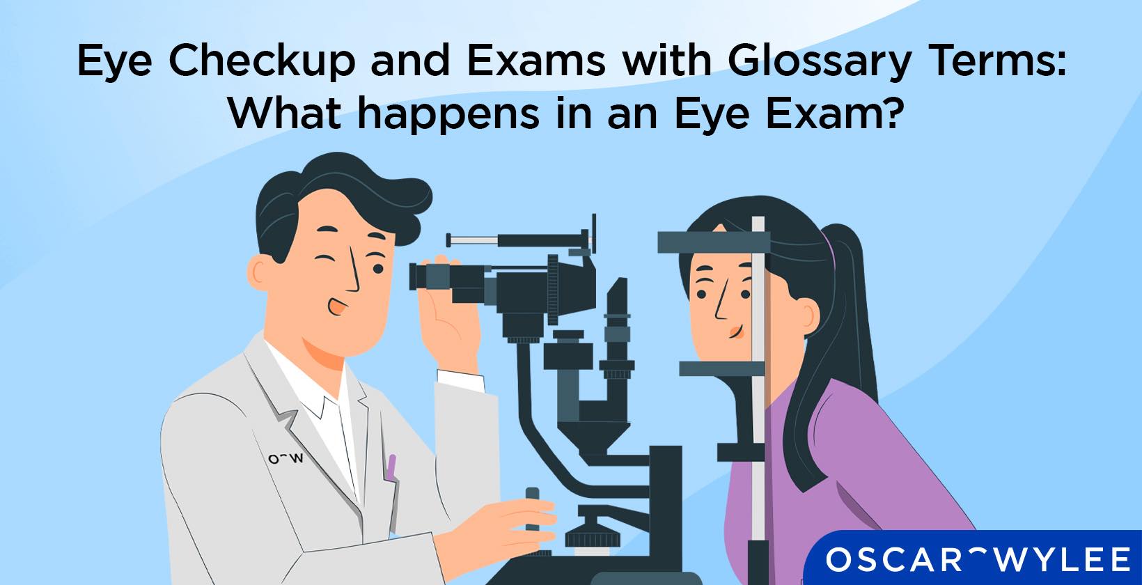

Eye Checkup and Exams with Glossary Terms: What happens in an Eye Exam?
An eye checkup and exam is an important aspect of taking care of your eye health and your overall health. It is performed by an eye care professional, most often an optometrist, and is a check of your vision and eyes.
Eye exams and checkups are necessary as they detect eye conditions or diseases that threaten the health of your eye and provide you with any vision correction needed. Eye exams are used to detect serious eye diseases such as glaucoma, cataracts, macular degeneration and diabetic retinopathy. For many eye conditions, early detection is a key aspect of the treatment and prevention of worsening conditions. Eye tests are most commonly performed by optometrists, but they are also done by ophthalmologists. Optical dispensers are trained to perform the pre-testing procedure. There are different eye tests within an eye exam that make up the procedure such as an eye pressure test which is conducted during pre-testing, a visual field test or a visual acuity test.
What Happens in an Eye Exam?
An eye exam is a procedure, most commonly performed by an optometrist, where a person’s eyes are checked to assess their vision. It is an important part of maintaining eye health as an eye test is used to detect a wide range of eye diseases and vision issues. The results of an eye test enable eye care professionals to provide tailored advice on how to best support your visual needs, which vary with age, lifestyle and the type of work you do.
An eye test begins with pre-testing, where an eye care professional will test the pressure of the eye and take a picture of the back of the eye (not all Oscar Wylee stores offer this machine). Afterwards, the patient will see the optometrist, who starts off the appointment by asking for information on their medical history as well as family medical history, why they are here, what concerns they have, any symptoms and any existing prescriptions.
Easily book an eye test with our dedicated Oscar Wylee optometrists on our online booking page. Using high-quality equipment, our eye tests take approximately 20 minutes, which is quick and convenient, meaning you can come in on your lunch break or on the weekend. The eye test experience at Oscar Wylee is an easy and enjoyable way to look after your eye health and with bulk billed eye tests for eligible Medicare card holders, there is no gap or out-of-pocket expenses.
What are the Types of Eye Exams?
There are four main types of eye exams that are performed for different purposes and are only necessary for certain people. According to Australia’s National Health Advice Service Healthdirect, the three main eye tests are a refraction test, an eye muscle test and an internal and external test. The definitions of these tests are listed below.
- Refraction test: A refraction test checks a patient's ability to see objects up-close and at a distance, to determine their prescription.
- Eye muscle test: An eye muscle test checks to ensure the muscles around the eye are working together correctly.
- Internal and external test: An internal and external eye test looks at the inside and outside of the eyes for any issues.
What is a Glaucoma Test for Eye Health?
A Glaucoma test for eye health is used to examine the eye for a condition known as glaucoma. Glaucoma is tested using many different procedures, although the most common is an eye pressure test which is performed during pre-testing. Glaucoma occurs due to a buildup of intraocular pressure and causes deterioration of the optic nerve. It begins as a loss of peripheral vision until it devolves into complete vision loss.
An optometrist may perform three tests to assess for glaucoma. The first is an eye pressure test which is a screening tool performed during pre-testing with an NCT (non-contact tonometer). Contact tonometry is also used to diagnose glaucoma. The next is an eye health check which includes an assessment of your optic nerve. The third test for glaucoma is a visual field test, where a person’s vision is mapped with a visual field machine, checking peripheral vision.
What Does Ophthalmic Test Mean?
An ophthalmic test is a series of comprehensive tests performed to check the health of your eye and vision. Ophthalmology exams are performed by either an ophthalmologist or an optometrist.
What is the Average Duration for an Eye Exam?
The average duration of an eye exam depends on many factors such as the tests performed or the specific patient and their needs. For example, if a person has more serious eye conditions, an eye test could take up to an hour but for a standard eye test, it is usually 20 to 30 minutes.
At Oscar Wylee, our eye test appointments are approximately 20 minutes, meaning they are quick and convenient.
How Often Should I Get My Eyes Checked?
At Oscar Wylee, we recommend that everyone should have an eye test with an optometrist at least once every 2 years. If you are over the age of 65, then an annual eye test is suggested. This frequency of eye tests depends on your health, age and if you have a risk of certain eye conditions. There are certain factors that require you to have more frequent eye tests.
Medical conditions: If you have medical conditions such as high blood pressure and diabetes, you may require more frequent eye tests as they may affect your vision.
Diagnosed eye conditions: For people with existing eye conditions such as glaucoma or retinal detachment, your eye care professional may recommend you come in for regular eye tests.
Age: The recommended frequency of eye tests changes depending on age. For example, if you are older you are more likely to develop certain eye conditions leading to more frequent eye tests.
What are the Procedures in an Eye Exam?
There are many different procedures that make up an eye exam, some common and some that are only performed for a patient with specific eye care needs. The procedures performed in an eye exam and their definitions are listed below.
- Visual Acuity Exam for Eyes: A visual acuity test measures the sharpness and clarity of your vision.
- Visual Field Test for Eyes: A visual field test is conducted to check your visual field which includes the sides of your vision.
- Pupillary Reactions for Eyes: Pupillary reaction tests assess optic nerve function using the pupil’s light reflex.
- Cover Test for Eyes: A cover test is used to determine misalignment in eyes and performed by an optometrist or ophthalmologist.
- Retinoscopy for Eyes: A retinoscopy measures the refractive error in a patient's eye to determine their prescription.
- Refraction for Eye Tests: A refraction test assesses a patient's ability to see at near and far distances.
- Slit Lamp Exam for Eyes: A slit lamp microscopically examines the eyes for problems or abnormalities.
- Pachymetry Tests for Eye: A pachymetry test measures the thickness of the cornea.
- External Exam for Eyes: An external exam inspects the alignment and position of the eyes as well as the pupils and extraocular movements.
- Retinal Examination for Eyes: A retinal exam, often known as funduscopy, evaluates the back of the eye including the retina.
- Eye Dilation Exams for Eyes: An eye dilation exam checks for various eye conditions such as glaucoma and diabetic retinopathy.
1. Visual Acuity Exam for Eyes
A visual acuity test is used to measure the sharpness and clarity of a person’s vision and how well they see at a distance. A visual acuity test involves a patient sitting across from a Snellen eye testing chart or a LogMAR chart set at 6 metres away. The optometrist instructs the patient to cover one eye and read letters that get progressively smaller until they are unable to read the letters clearly. This process is then repeated with the other eye covered. The further down the chart a patient sees, the better the visual acuity is. The lowest line they are able to read to is expressed as a fraction and is then used to describe visual acuity. Visual acuity is an important part of determining the clarity of your vision but it is only one aspect of a comprehensive eye exam.
2. Visual Field Test for Eyes
A Visual Field Test (VFT) for eyes is used to quantify how well a person sees through each eye individually and binocularly. It maps a patient’s visual scope and peripheral vision and helps identify vision loss and associated eye conditions. This test helps the optometrist establish a baseline and diagnose ocular injuries or diseases, including glaucoma.
A visual visual field test frame varies on the model of the machine and the test that is being done, from 10-40 minutes. The test involves identifying lights or objects that appear on the sides of your vision; this could be through a machine or performed by an optometrist or a trained team member. For this test, a patient is positioned on the machine with one eye covered. Fixating on the central light in the machine, the patient presses their response button each time they see a light pop up, using their peripheral vision. The lights vary in position, speed and brightness. The results are printed as a map and interpreted by the optometrist.
3. Pupillary Reactions for Eyes
Pupillary reactions for eyes, refers to how the pupil of the eye responds to light stimuli. The pupillary light reflex is an important part of eye health as it is used to assess optic nerve function. In a 2022 article in the National Library of Medicine, the function of a pupillary light reflex test is described to evaluate and manage abnormal pupillary light reflexes. This test is conducted using a penlight where the eye care professional will shine the light into one eye and then withdraw it for a few seconds and observe the pupils’ reactions.
4. Cover Test for Eyes
A cover test for eyes is used to determine the magnitude and type of ocular misalignment, also known as strabismus. According to the American Academy of Ophthalmology, a cover test is typically performed by optometrists and ophthalmologists using a sheer or opaque occluder that covers one eye. The patient fixates on a target and the eye care professional observes the shift in fixation in the non-occluded eye.
5. Retinoscopy for Eyes
Retinoscopy for the eyes refers to a testing technique that measures the refractive error in an eye. A refractive error occurs when the light entering the eye does not properly hit the retina. This could mean light hits before the retina, or behind the retina, resulting in vision issues. A retinoscopy helps to identify and determine the degree of the refractive error.
Optometrists use a retinoscope, a handheld tool that they look through, in conjunction with the retinoscopy technique, to determine a refractive error. According to the American Academy for Paediatric Ophthalmology and Strabismus, the retinoscope has a beam of light that shines from it, that your optometrist will direct onto the eye. The optometrist uses the retinoscopy technique to assess how this light bounces off the retina and determines the refraction issue.
6. Refraction for Eye Tests
A refraction test for the eyes is performed by an eye care professional to determine the necessary correction for a refractive error, according to the non-profit organisation, My Vision. A refractive error is a category of visual issue that affects a patient’s ability to see clearly. Refractive errors include myopia, hyperopia and astigmatism. The most common tests for eye refraction are retinoscopy, autorefraction and phoropter refraction.
Retinoscopy: Using a retinoscope, the optometrist will move the light across both eyes to determine how the light reflects off the retina.
Autorefraction: This test involves an autorefractor to measure refraction, which is a computer-controlled machine.
Phoropter refraction: Uses a phoropter to measure refraction which is a device that looks like a large mask and a patient looks through a combination of lenses.
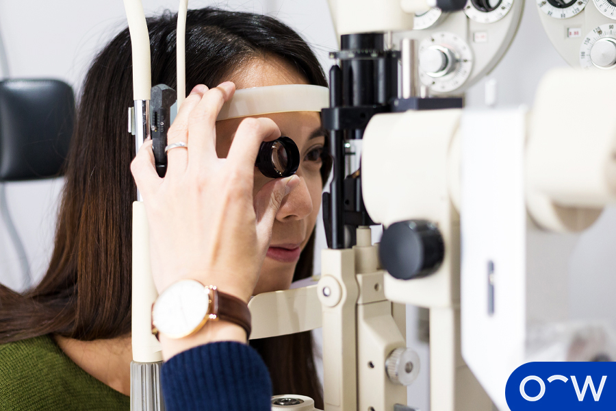
7. Slit Lamp Exam for Eyes
A slit lamp exam for the eyes refers to a test in which a microscope with a bright light is used to examine the various structures of the eyes such as the cornea, the lens, the optic nerve and the retina. Your eyes may be dilated for this procedure. According to the American Academy of Ophthalmology, a slit lamp test is used to observe parts of the eyes for abnormalities and eye diseases such as glaucoma, macular degeneration and cataracts.
To conduct a slit lamp test, an optometrist will position the patient’s chin in a chin rest connected to the microscope and the forehead rests on a band, also connected to the microscope. The optometrist then uses the beam of light from the microscope to inspect the eye.
8. Pachymetry Tests for Eye
A pachymetry test for the eyes is used to measure the thickness of the cornea. It is an important aspect of eye health as it can detect if a patient’s cornea is swollen, which can be caused by certain eye conditions or even by wearing contact lenses. A pachymetry test is performed in two ways, using an ultrasound or an optical pachymeter.
9. External Exam for Eyes
An external exam for eyes is defined by Frank C. Bell in a paper published in the National Library of Medicine, as an external eye examination involving the external eye structures including the eyelids and surrounding tissues, the cornea, conjunctiva, anterior chamber and the lacrimal apparatus. The purpose of an external eye exam is to gain information about the patient that could help diagnose certain eye conditions or diseases. A pocket flashlight or more commonly a slit lamp is used in an external eye exam to inspect areas of the eye such as the cornea and the anterior chamber.
10. Retinal Examination for Eyes
A retinal examination for the eyes, also known as a funduscopy, allows an eye care professional to evaluate the back of a person’s eye. This includes looking at the retina and optic disc. A retinal exam often involves a person’s pupils being dilated for better visualisation for the optometrist and using an ophthalmoscope which shines a beam of light into the pupil in order to examine the back of the eye.
11. Eye Dilation Exams for Eyes
An eye dilation exam for eyes, refers to a procedure in which an optometrist dilates your eye to get a better view of its inner structures. This exam helps an optometrist to get a more in-depth picture of your eye health, beyond a standard eye exam. The exam is used to help check for eye conditions such as glaucoma, macular degeneration and diabetic retinopathy.
An optometrist conducts an eye dilation exam by administering eye drops. These eye drops will dilate, or widen, your pupil so the optometrist can see the inner structures of the eye more clearly. Once the pupil is dilated, the optometrist may complete more eye tests such as visual acuity testing.
What You Need to Know About Eye Anatomy to Understand Eye Exams and Checkups?
It is important to know about eye anatomy in order to understand eye exams and checkups. The eye (oculus–Latin term for eye), approximately 2.5 centimetres in diameter, is one of the most important organs in the body. It processes the light that is reflected off surfaces into electrical signals creating the images we see, allowing people to interpret shapes, faces, colours and depth. The eye rests in a socket in the skull and is surrounded by 6 motion-regulating muscles and many layers of fatty tissue for protection and flexibility. There are many different parts of eye anatomy and various procedures, surgeries and treatments to address eye conditions and diseases.
An eye exam tests the function of your vision and assesses for any eye conditions or diseases using various tools such as a slit lamp or a DRS machine (digital retinal scanning).

1. Applanation Tonometry
Applanation tonometry is a procedure that tests the pressure in a person's eye. Eye pressure is an important aspect of your eye health as high eye pressure may potentially damage your optic nerve and cause glaucoma. For this exam, eyes are numbed using drops with dye in them and the patient’s head is positioned on a slit lamp, a diagnostic tool used in ocular examinations. The eye pressure is measured using a small tip that gently touches the eye's surface.
2. Achromatopsia
Achromatopsia is an eye condition that causes complete or partial absence of colour in a person’s vision meaning they only perceive white, black and shades of grey, no colour. According to the National Library of Medicine, someone with achromatopsia may have a sensitivity to glare and light, and have a significantly diminished sharpness of vision.
Achromatopsia is different from colour blindness as a person who is colour blind is able to perceive colour but has trouble telling the difference between certain colours.
3. Aqueous humour
Aqueous humour is a part of the eye anatomy and is located in the anterior and posterior chambers of the eye. It is a transparent, thin fluid that is made up of almost completely water with a small number of proteins and other nutrients. The aqueous humour’s main function is to supply nutrients to the lens and cornea.
4. Choroid
The choroid is a tissue layer that sits in between the sclera and the retina. According to the National Cancer Institute, it provides oxygen and nourishment to the outer layers of the retina and maintains the volume and temperature of the eye. The choroid functions to shield the eye from harmful reflection and light. If the choroid is damaged, the retina may bleed and the damage eventually results in the loss of peripheral vision.
5. Choroiditis
Choroiditis, also known as Serpiginous Choroiditis, is a rare eye disorder that causes recurrent lesions that affect two layers of the eye, the retinal pigment epithelium and the choriocapillaris. There are commonly no symptoms of this eye disorder unless the macula is damaged. Choroiditis most often affects both eyes and the cause of this eye disorder is unknown.
6. Conjunctiva
The conjunctiva is part of the eye anatomy and is a protective, clear membrane that covers the inside of a person's eyelid as well as the sclera. Its purpose is to secrete fluids that keep the eyes lubricated and protected from infections and outside bacteria and bodies, such as dust. The conjunctiva is defined by Mayo Clinic as creating the mucus layer that forms part of a person’s tears. The conjunctiva can be affected by eye disorders such as conjunctivitis, also known as pink eye, and a subconjunctival haemorrhage.
The conjunctiva comprises three aspects, the bulbar conjunctiva, the palpebral conjunctiva and the fornix conjunctiva. These three layers ensure that no foreign bodies or objects travel behind the eye.
7. Conjunctivitis (Pink Eye)
Conjunctivitis, also known as pink eye, is an inflammation of the conjunctiva, a lining covering the white part of the eye and underneath the eyelids. The small blood vessels within the conjunctiva dilate and cause the eye to take on a pinkish-red colour (that is where the term “pink eye” comes from).
The most common causes of conjunctivitis are allergies, bacterial infections, viral infections and contact lens wear. Besides pink eye, there are other associated conjunctivitis symptoms such as watery eyes, crusting in the corners of the eyes, white, yellow or stringy discharge and itchy, irritated, sore, and gritty feeling in the eyes.
8. Cornea
The cornea is the clear front surface of the eye, acting like a transparent window according to the National Cancer Institute. The cornea has a refractive index and it covers the pupil and the iris, allowing light to enter the eye. The cornea functions by bending and refracting light that the eye receives which is achieved by the pupil changing size, becoming smaller or larger, depending on the volume of light. The cornea protects the eye by filtering out harmful glare from the eye, such as ultraviolet rays from the sun. The cornea does not contain blood vessels, rather it is composed of proteins and cells, this means it receives its nutrients from tears and the aqueous humour, which is a fluid similar to plasma.
9. Cyclitis
Cyclitis is a type of uveitis, which is an inflammation of the tissue within the eyeball. Cyclitis is a type of anterior uveitis and causes inflammation to occur in the muscles that operate the lens.
10. Dilation
Dilation occurs when the pupil, which is the black centre of the iris, is enlarged. Pupils change their width depending on the eye’s exposure to light, for example when they are exposed to bright light the pupil will constrict. When a person is in a dark room, the pupils enlarge, which allows them to see in low lighting.
11. Dilated Pupillary Exam
A dilated pupillary exam is performed on patients to check their eye health as it can detect certain eye diseases. This painless and simple exam involves an eye care professional administering eye drops to dilate the pupil in order to check for eye diseases. According to the National Eye Institute, a dilated pupillary exam checks for diseases such as glaucoma, age-related macular degeneration and diabetic retinopathy.

12. Fluorescein Angiography
Fluorescein angiography is a procedure in which an ophthalmologist uses a special camera to take images of the retina. These images allow the eye doctor to view the blood vessels and other aspects that exist in the back of the eye.
The American Academy of Ophthalmology reports that fluorescein angiography is used to detect and diagnose eye conditions and diseases such as macular degeneration, macular edema, diabetic retinopathy and ocular melanoma.
13. Hyperopia
Hyperopia, also known as farsightedness, is a refractive error in which images are focused behind the retina rather than on it, making it hard to see objects at a near distance. Some people with hyperopia may struggle with both long and short distances.
Hyperopia is corrected with a converging or plus power lens. Common causes of hyperopia include a decrease in refractive index (cortical cataracts), the eye is shorter than normal and the cornea is flatter than normal. Hyperopia is different from presbyopia because with presbyopia, the trouble with near vision is due to the decreased lens flexibility that comes with age.
14. Intraocular
Intraocular is a term that refers to anything occurring, implanted or administered by entering the eyeball. It is often associated with intraocular pressure which refers to the fluid pressure of the eye.
15. Iris
The iris is a part of the eye anatomy and makes up the coloured portion of the eye. It contains muscles that contract to adjust pupil size depending on light conditions. The iris is constantly changing its dilation due to how much or little light there is which allows the eye to see clearly. The iris is as unique as a fingerprint, meaning no two people have the same eye colour.
16. Low Vision
Low vision is an eye problem that affects your ability to do everyday activities like reading and driving. Low vision is caused by various eye conditions such as cataracts and age-related macular degeneration and cannot be corrected with glasses.
According to the National Eye Institute, there are four main types of low vision. These include blurry or hazy vision, central vision loss, peripheral vision loss and night blindness.

17. Macula
The macula is the part of the eye anatomy that processes what you see in your central vision, that which is directly in front of you. It is found at the back of the eyeball. The macula functions to help a person understand specific details and parts of images such as colour, and text on a page and discern the difference between faces.
18. Macular Oedema
Macular edema occurs when there is swelling in the part of the retina which causes blurry or wavy vision as well as dull colour vision. According to the National Eye Institute, the most common cause of macular edema includes diabetic retinopathy. Other causes include uveitis and age-related macular degeneration.
19. Myopia
Myopia, also known as nearsightedness, is a refractive error that occurs when light focuses before the retina rather than on it due to curvature (increased corneal curve) or axial (increased eyeball length). It causes patients to have difficulty seeing distant objects clearly. Treatment may include glasses, contact lenses, laser or IOL surgery. A minus-power lens is prescribed to bring the light back to focus on the retina.
Myopia is one of the most prevalent eye conditions on the planet. It is estimated that by 2050, nearly 5 billion people will be affected by myopia, according to a 2016 study by the American Academy of Ophthalmology.
20. Nyctalopia (night blindness)
Nyctalopia is a term that refers to a person who has difficulty seeing in dim light or at night. People with nyctalopia will usually find their daytime vision is unaffected, according to an article published in the National Library of Medicine, by Divy Mehra and Patrick H. Le. Night blindness is caused by the eye’s inability to respond quickly from lightness to darkness.
21. Non-Contact Tonometry
Non-contact tonometry (NCT), also known as air puff tonometry, uses a rapid air pulse that flattens your cornea. The period when your eye becomes flat, is how the pressure is measured, which is derived from the monitored reflection of an incident infrared light. The NCT method tests your intraocular eye pressure which is the internal pressure of the eye. It is part of the pre-testing routine and can be built into an autorefractor.
22. Ocular
Ocular refers to anything that is perceived or done by the eye or relates to the eye in any way.
23. Ophthalmologist
An ophthalmologist is a medical doctor that specialises in eye care and provides advanced visual care that your optometrist cannot. This includes assessing and managing serious eye conditions like advanced glaucoma and performing surgeries.
As medical doctors that are specially trained in eye health, ophthalmologists are qualified to carry out a variety of eye care services such as the treatment and management of complex eye diseases and performing eye surgery. Ophthalmologists receive patients from referrals, either by a General Practitioner or from optometrists who have noticed serious eye conditions beyond their expertise. Ophthalmologists then assess the patient and create treatment plans for their eye or vision problems.
24. Ophthalmoscope
An ophthalmoscope is a tool used by eye care professionals that allows them to look at the back of the eye. An ophthalmoscope uses a beam of light to assess the health of the optic nerve, retina, vitreous humour and vasculature.
25. Optic Nerve
The optic nerve transmits all visual information from the retina to the brain, to process the sensory information so that a person can see. The optic nerve is an extension of your central nervous system along with the brain and the spine.
26. Optometrist
An optometrist is the most common type of eye care professional and the one you are most likely to encounter when having an eye exam. They are eye care professionals who have completed a university degree in optometry. Optometrists provide comprehensive eye exams which address both visual concerns relating to prescription errors as well as ocular surface diseases. Your optometrist is your first point of contact in your eye health journey.
Most optometrists in Australia provide services such as complete eye exams, identification and management of vision problems and eye infections, prescription of eyeglasses, diagnosis and treatment of eye disorders, providing advice on eye care and referring patients to ophthalmologists as needed.
27. Peripheral Vision
Peripheral vision is the part of your sight at the sides of the visual field, as opposed to what you are able to see straight in front of you. Your peripheral vision is tested in a visual field test and used to detect eye conditions such as glaucoma.
28. Photocoagulation
Laser photocoagulation is a type of laser surgery performed on the eyes to treat age-related macular degeneration, an eye disease that can result in vision loss. The surgery involves the doctor using a laser to burn away abnormal structures in the retina or to deliberately scar the eye.
29. Presbyopia
Presbyopia is the inability to see objects at close distances. The refractive error presbyopia occurs naturally with age as the protein composition of the crystalline lens changes, reducing its flexibility. Light focuses behind the retina meaning things need to be further away to see. Presbyopia is corrected with an ADD power in reading glasses or multifocals glasses.
30. Pupil
The pupil is the centre of the iris where light enters. It works like a camera aperture, if the light is bright, the pupil is smaller, and if it is darker, the pupil is dilated.
31. Refraction
Refraction is described as the bending of light rays as they pass from one object to another, according to The National Eye Institute. In the eye, refraction occurs as the lens and cornea bend the light to focus them on the retina.
32. Refractive Error
A refractive error is a type of vision issue that affects a person’s ability to see clearly. They are the most common type of vision problem and occur when the shape of the eye stops light from focusing correctly on the retina.
There are four different types of refractive errors defined by the National Eye Institute.
Farsightedness: Also known as hyperopia, farsightedness causes near objects to appear blurry.
Nearsightedness: Also known as myopia, nearsightedness makes distant objects look blurry.
Presbyopia: Presbyopia is an age-related eye condition that makes it difficult for older people to see things close up.
Astigmatism: Astigmatism makes both far-away and nearby objects appear distorted or blurry.
33. Retina
The retina is a fine layer of nerve cells at the back of the eye, responsible for analysing colour, intensity and form, like the film in a camera. The retina receives visual information through millions of light-sensitive cells and sends it to your brain via the optic nerve, allowing you to see.
34. Retinal Tomography
Retinal tomography is used to precisely observe and document the optic nerve head. This diagnostic procedure is essential in managing and diagnosing glaucoma. According to the University of British Columbia, the procedure involves taking images of the optic nerve and surrounding retina to create a 3D image of the optic nerve.
35. Retinitis Pigmentosa
Retinitis pigmentosa (RP) is the name for a group of rare eye diseases affecting the retina by causing cells to slowly break down over time which leads to vision loss. According to the National Eye Institute, retinitis pigmentosa is a genetic disease that people are born with and there is no cure, however, vision rehabilitation programs are available. Although there are certain Australian institutes researching gene therapy as a cure for retinitis pigmentosa
The symptoms of RP are loss of peripheral vision, difficulty adjusting to dim lighting, tunnel vision, loss of colour vision and sensitivity to light.
36. Sclera
The sclera is the white part of the eye. The sclera is described as an opaque, fibrous, protective outer layer of the eye and contains collagen and elastic fibre.
37. Strabismus
Strabismus also known as eye misalignment, is a muscle imbalance that causes double vision, unstable images, eye strain and fatigue, as well as reduced depth perception and 3D vision. Strabismus occurs when the eyes struggle to work together and converge images. A person with strabismus will have eyes that have a turn.
38. Tunnel vision
Tunnel vision, also known as peripheral vision loss, occurs when a person loses their peripheral sight (side vision). Tunnel vision means a person is still able to see clearly out the front of their vision but may have gaps or holes out the sides of their visual field.
39. Ultrasound
An ultrasound is used to assess the structural integrity of the eye and also diagnose conditions such as tumours and retinal detachment. An ultrasound works by reflecting sound waves to create an image that provides information that cannot be obtained by direct visualisation.
40. Visual Acuity
Visual acuity is defined as a person’s ability to see at a distance, measuring the sharpness and clarity of their vision. It is tested during a routine eye exam by an optometrist using a Snellen Chart, testing one eye at a time. Your visual acuity is important as it is a consistent way for an eye health professional to detect changes in your vision.
Visual acuity is denoted using fractions. The normal measure of visual acuity is 20/20 vision, or 6/6 when using the metric system. If a person has 20/20 vision, it means they see objects clearly from 20 feet away.
41. Visual Field
Visual field describes the total area in which objects can be seen in the sides (periphery) of their vision when they focus on a central point. A visual field test is conducted by an eye health professional to measure the amount of vision a patient has in each eye.
Certain medical conditions can cause people to experience loss of their peripheral vision such as diabetes and high blood pressure, this is why visual field testing is important as it monitors the effect of these medical conditions on a person’s vision.
42. Vitrectomy
A vitrectomy is a type of eye surgery performed by an ophthalmologist to treat different problems with the vitreous and retina. A Vitrectomy is conducted if a patient has eye conditions such as a macular hole or pucker, diabetic retinopathy, a severe eye injury or an infection in the eye.
The surgery involves the ophthalmologist making a small incision in the sclera, the white of the eye. The doctor may do one or more of the following steps: remove cataracts, take away scar tissue on the retina, get rid of cloudy vitreous and remove any foreign objects from the eye.
43. Vitreous humour
Vitreous humour is a clear, gelatinous mass that fills the space in the eye between the lens and the retina. It is composed mostly of water and functions to ensure vision clarity, maintain the shape of the eye and absorb impact to the eye or head.
According to the Vision Eye Institute, over time, vitreous humour may shrink and liquefy and the collagen and protein can become stringy which potentially causes floaters in the eye.
How to Perform Eye Vision Tests at Home?
It is not recommended to perform eye vision tests at home as they are not reliable and often provide inaccurate results. It is best to have an eye test performed by a professional who has the knowledge, experience and equipment to provide you with the most accurate results.
If you need an eye test of any kind, it is best to book an appointment with an optometrist. They perform the eye vision test and interpret the results correctly.
Why is Understanding Eye Exams important?
It is important to understand eye exams because they are a crucial aspect of your eye care and overall health. By learning the different tests performed and possible eye conditions, it allows you to know why eye exams are beneficial and should not be ignored.
Who Can Perform Medical Eye Tests?
There are two main eye care professionals who can perform eye tests, these include optometrists and ophthalmologists, and there are also certain procedures an optical dispenser can conduct. The definition of these professions and their duties are listed below.
- Optometrist: An optometrist is the person you encounter when you book an eye test as it is one of their main roles. They provide comprehensive eye exams which address both visual concerns relating to prescription errors as well as ocular surface diseases.
- Ophthalmologist: An ophthalmologist is an eye doctor who performs surgeries and provides specialised care for eye conditions that cannot otherwise be managed by optometrists. You see an ophthalmologist if you have been referred by an optometrist, General Practitioner or medical specialist.
- Optical Dispenser: The role of an optical dispenser is to interpret the prescriptions given to them by the optometrist, with some performing pre-testing. Optical dispensers take pupillary distance measurements and ensure eyewear fits comfortably and correctly on a patient.
What is the Cost of a Medical Eye Test?
In Australia, Medicare provides a subsidy for Australians to regularly get their eyes tested. For eligible Medicare cardholders, people receive a bulk billed eye test with no gap or out-of-pocket expenses. If you are not covered under Medicare, eye tests at Oscar Wylee are $70 each.
Medicare is Australia’s healthcare system that officially started in 1984. Since then, it has provided coverage and gap payments for Australian citizens and permanent residents for all payments going towards health costs and medical aid. According to Services Australia, bulk billing occurs when a healthcare specialist sends the entirety of the bill for their Medicare-covered services to the government to be paid in one instance.
Are you Obligated to Buy Glasses if You Have an Eye Test?
No, you are not obligated to buy glasses after you have had an eye test. An eye test is a way to check your eye health and is an important part of eye care. Your optometrist may prescribe that you wear glasses to correct your vision although it is your decision to listen to their advice or not.
We recommend that if you are prescribed glasses by an eye care professional, if you are able to, you purchase these corrective devices. Prescription glasses improve your day-to-day life and are safer, especially for driving.
Does Oscar Wylee Provide Free Optometrist Tests?
At Oscar Wylee, a qualified optometrist is available in-store to conduct a bulk billed eye test for all eligible Medicare cardholders. An Oscar Wylee eye test has no gap and requires no out-of-pocket expenses necessary however certain driver's license testing and occupational testing are not covered. Regardless of Medicare status, anyone seeking a professional eye test is welcome at Oscar Wylee stores.

What are the Statistics of Medical Eye Tests?
Optometry Australia reported that in 2019, Australian optometrists performed a record of more than 10 million eye examinations under Medicare. In 2019, Optometry Australia found that the number of registered optometrists in Australia has exceeded 6,000.
A study conducted by the Australian Bureau of Statistics in 2017-18 found that in Australia, over 13 million people have one or more long-term chronic eye conditions. This includes:
- 7.2 million with hyperopia (long-sightedness)
- 6.3 million with myopia (short-sightedness)
- 1.4 million with astigmatism
- 687,000 with presbyopia (loss of focusing ability with age)
- 549,000 with colour blindness
- 411,000 with cataracts
- 244,000 with macular degeneration
- 131,000 with blindness (complete and partial)
Also, according to the Australian Government Department of Health and Aged Care, around 90% of all vision impairment and blindness in Australia are treatable or preventable if detected early.