 1300 164 990
Visual Field Test: How Does it Work, Types and Benefits
Published on June 7th, 2024
Updated on February 13th, 2025
 Australia

A visual field test (VFT) is used to quantify how well a person can see through each eye individually. A visual field test helps the optometrist establish a baseline of vision and diagnose ocular injuries or diseases, including glaucoma. A visual field test, also known as a peripheral vision test, involves identifying lights or objects that appear in the sides of a person’s vision (periphery); this could be through a machine or performed by an eye care professional. The different types of visual field tests include confrontation visual field test, Amsler grid, static automated perimetry test, kinetic perimetry visual field test and frequency doubling perimetry. Keep reading to learn more about how a visual field test works and its benefits.
What is Visual Field Testing?
Visual field testing is a non-invasive examination that is useful in the diagnosis and monitoring of conditions such as glaucoma as well as neurological conditions such as pituitary tumours. A visual field test is performed to quantify how well a person can see with each eye individually. It is most often performed using a visual field machine which is a specialised piece of equipment that records how well each eye can see, and based on the results can reveal links to neurological conditions. A visual field test aids an optometrist in establishing a baseline for a person’s vision to diagnose eye diseases or injuries. The results of a visual field test can be used to monitor progressive conditions that affect a person's peripheral visual field and/or central visual field. The results of a visual field test will typically be printed as a map and interpreted by the eye care professional administering the test.
Why Do You Need a Visual Field Test?
A visual field test is beneficial for people who have progressive eye diseases and neurological conditions affecting their visual fields as well as drivers who need to fit certain criteria to continue driving. Visual field testing can help detect early signs of certain eye diseases such as glaucoma and should be had as often as an optometrist recommends. Visual field tests are important as they can detect problems with a person’s field of vision that are otherwise difficult to notice. If a person does have deficiencies in their field of vision such as a blind spot, their brain may be filling these in, leaving them unable to identify any changes in eyesight until it is too late. Visual field testing can also detect conditions affecting visual pathways between the optic nerve and the visual cortex. These conditions primarily include tumours.
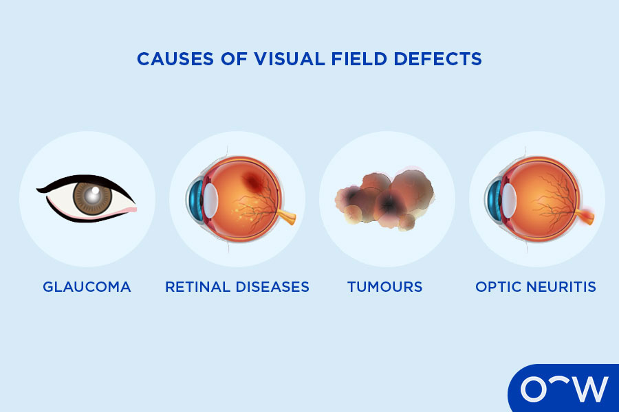
What Causes Visual Field Defects?
A visual field defect is anything that damages the pathways that carry visual signals to the back of the eye and then to the occipital lobe, which is the brain’s visual processing centre, according to Barrow Neurological Institute. The causes of visual field defects include glaucoma, retinal diseases, tumours and optic neuritis. These visual field defects and their definitions are listed below.
- Glaucoma: Glaucoma is a very common cause of visual field defects. Glaucoma is the term for a group of eye diseases that occur when the optic nerve is damaged. It can cause irreversible low vision and loss of vision, most commonly in a person's peripheral field.
- Retinal diseases: Retinal diseases refer to eye conditions that affect the retina. Common symptoms of retinal diseases include blind spots in a person’s central or peripheral vision. Retinal diseases include retinal detachment and tears, diabetic retinopathy and retinitis pigmentosa. Retinal diseases are often diagnosed in a retinal exam.
-
Tumours: Tumours may cause visual defects as they can press on the optic
nerve leading to damage, according to the American Academy of
Ophthalmology. Tumours are considered a central nervous system
problem.
- Optic neuritis: Optic neuritis is an eye condition characterised by damage to the optic nerve due to inflammation. Optic neuritis can be detected using a kinetic perimetry visual field test as it is used to find vision problems found in the central nervous system, according to Very Well Health.
What are the Different Types of Visual Field Tests?
The different types of visual field tests are confrontation visual field test, Amsler grid, static automated perimetry test, kinetic perimetry visual field test and frequency doubling perimetry. These visual field tests and their definitions are listed below.
- Confrontation visual field test: A confrontation visual field test is a common visual field test that does not involve machines. The eye care professional will test peripheral vision by holding objects in a patient’s periphery and asking if they can see them.
- Amsler grid: An Amsler grid is a basic visual field test using a grid of squares with a large dot in the middle. While looking at this dot, a patient is asked to describe if any areas look blurry, wavy or distorted.
- Static automated perimetry test: A static automated perimetry test involves the patient looking at a bowl-shaped instrument called a perimeter where flashing lights will appear on the sides of their vision. The patient is asked to press a button when they see any of these lights.
- Kinetic perimetry visual field test: In a kinetic perimetry visual field test, the patient is shown moving light targets in a visual field test machine and asked to identify when they are visible in their periphery. Kinetic visual field testing is used to map the complete visual field.
-
How Does a Visual Field Test Work?
A visual field test involves being positioned in front of a machine that shows flashing lights of differing intensities in which the patient will record their responses by clicking a button. Each patient's eye will be tested separately with the opposite eye covered by an eye patch. During the test, there will be some spots that are not visible which is normal and part of the exam to help generate reliable results.
Who Performs a Visual Field Test?
An eye care professional, such as an optometrist or ophthalmologist, will perform a visual field test. An optometrist is the most common eye care professional that will perform a visual field test. A visual field test may be performed as part of a standard eye test by an optometrist who will also test a patient’s visual acuity and eye health, among other tests. An ophthalmologist, also known as an eye doctor, may also perform a visual field test.
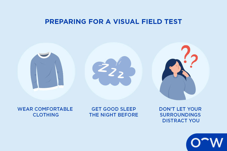
How to Prepare for a Visual Field Test?
There are certain things a person can do to prepare for a visual field test. To ensure the results of the visual field test are as accurate as possible and to prevent the patient from having to redo the test, they should wear comfortable clothing, get a good night’s sleep the night before and not let their surroundings distract them. These ways to prepare for a visual field exam are listed below.
- Wear comfortable clothing: It is important to wear comfortable clothing when preparing for a visual field test as the more comfortable the patient is, the smoother the process will be.
-
Get a good night’s sleep the night before:
A good night’s sleep before the appointment can be very beneficial
as if the patient is tired in the visual field test, it could affect
the results.
- Don’t let your surroundings distract you: Where possible, a person should try not to let their surroundings be a distraction as it is best to be fully focused on the visual field test to provide accurate results.
How Long Does a Visual Field Test Take?
How long a visual field test takes can vary from person to person and also depends on the type of visual field test being performed. On average, a visual field test can take between 10 and 40 minutes. If the test results are inconclusive, the visual field test will be repeated, likely on another day.
How Much Does a Visual Field Test Cost?
The cost of a visual field test varies depending on where the procedure is done. A visual field test at Oscar Wylee can be bulk billed with Medicare, otherwise the cost is $70.
What are the Benefits of a Visual Field Test?
There are many benefits to a visual field test as it is a crucial aspect of testing a patient’s eye health. The benefits of a visual field test include diagnosing eye diseases, identifying neurological conditions and assessing a person’s fitness to drive. These benefits of a visual field test are listed below.
- Diagnosing eye diseases: A visual field test is a crucial procedure performed to identify vision loss and associated eye conditions. Glaucoma is one of the main eye conditions that can be diagnosed using a visual field test.
-
Detect neurological conditions:
A visual field test can help detect neurological conditions such as
pituitary tumours, which are unusual growths that develop in the
pituitary gland, according to the Mayo Clinic.
- Assess a person’s fitness to drive: A visual field test is part of the assessment to determine whether a person is fit to hold a driver’s licence. Alongside the visual field, other measures for a person’s fitness to drive include their visual acuity, colour vision and diplopia.
How do I Read My Visual Field Test Results?
There is no need for the patient to read visual field test results as the results will be interpreted and communicated by the optometrist or ophthalmologist administering the test. According to Very Well Health, the results of a visual field test, also known as a peripheral vision test, are often presented in charts and may include a decibel scale, grey-scale map, mean deviation, total deviation and pattern deviation. If a patient has abnormal visual field test results, they may be referred to an eye specialist for further tests or treatment.
How do Visual Field Tests Help Detect Glaucoma?
Visual field tests are crucial in diagnosing and treating glaucoma, according to Glaucoma Australia. A visual field test, also known as a peripheral vision test, is used to diagnose glaucoma, as one of its main symptoms is a loss of peripheral vision. A visual field test examines a patient’s peripheral vision by having them identify lights to determine if they have blind spots in their eyesight. According to the BrightFocus Foundation, visual field tests are repeated periodically to track the progression of glaucoma to see if it is getting worse or is stable.
How can Visual Field Tests Prevent Further Vision Loss?
Visual field tests can prevent further vision loss by detecting eye conditions so the patient can begin treatment. Early detection of eye conditions is extremely important as if proper treatment is started, it can prevent vision loss and blindness in many eye conditions. It is important to have regular eye tests so an optometrist can assess your eye health and perform a visual field test if they deem it necessary.
Read Visual Field Test: How Does it Work, Types and Benefits in other Oscar Wylee regions and their languages.
Australia