 1300 164 990

Retinal Exam: Importance, How it Works, and Benefits
Published on May 1st, 2024
 Australia


A retinal exam, also known as an ophthalmoscopy or fundoscopy, enables the optometrist to examine the back of the eye including the optic disc and the choroid with an increased depth of focus. The three types of retinal exam are a direct examination, indirect examination and a slit-lamp examination and all typically involve the use of magnification and a bright source of light. An optometrist may specifically order a retina test to check for certain conditions that can affect the blood vessels such as optic nerve damage, retinal tear or detachment, glaucoma, retinal infections, macular degeneration, melanoma, hypertension or diabetes.
Why is a Retinal Exam Important For the Retina?
A retinal exam is important for the retina as it enables the optometrist to visually assess the retina in full detail. The retina is defined as the light sensitive layer located at the back of the eye and helps us process the light that enters the eye into electrical signals. The brain can then interpret these electrical signals as visual images. A retinal exam is normally performed during standard eye tests, therefore, it is important to stay on top of your regular eye tests to ensure an eye care professional can identify any possible changes to the structures in your eye or your vision.
How Does a Retinal Exam Work?
A retinal exam works by dilating your pupils to help widen them and allow the optometrist to get a better view of the retina. However, retinal exams can be done with undilated pupils as well and may only require dilation if you are diabetic or have small pupils. During a retinal exam the optometrist may administer eye drops to dilate the pupil allowing more light to enter the eye. Each eye is examined using a specific magnifying lens that supplies a clear image of the vital tissues located at the back of the eye including the optic disk and the choroid. However, the process of the retinal exam may slightly differ depending on the type that needs to be performed.

What Techniques are Used in a Retinal Examination to See the Back of the Eye?
The techniques used in a retinal examination to see the back of the eye include a direct examination, a slit lamp examination and an indirect examination. Using a retinal exam as part of your regular eye test is how optometrists examine the deeper parts of the eye and first identify any warning signs of potential eye conditions. The techniques used in a retinal examination to see the back of the eye are listed below.
- Direct Examination: A direct examination simply involves the use of a handheld tool known as an ophthalmoscope to examine the back of your eye.
- Slit Lamp Exam: A slit lamp exam may require you to sit in a chair with a specific instrument positioned in front of you to then place your chin and forehead on. The optometrist will then look through a lens in combination with the microscope part of the instrument to view your retina.
- Indirect Examination: An indirect examination may either have you lying down or in a semi-reclined position. The optometrist will then shine a bright light in your eye with an instrument worn on their head and look through a lens that is held near your eye.
1. Direct Examination
A direct examination is a type of retinal examination that mainly involves the optometrist using a handheld tool, which is known as an ophthalmoscope. This tool has a light and different lenses that allows the optometrist to take a closer look at the back of the eye.
2. Slit Lamp Exam
A slit lamp examination will require you to be seated so you can position your chin and forehead on the slit lamp biomicroscope. The optometrist will then use the microscope on the machine together with a lens to examine the back of your eye. This method provides the same amount of view of the eye as an indirect examination does, but with increased magnification and narrower field of view.
3. Indirect Examination
An indirect examination may have you either lying down or in a semi-reclined position for the optometrist to then shine a bright light in your eye with an instrument worn on their head. The instrument is called a binocular indirect ophthalmoscope, it resembles a miner’s light and allows the optometrist to look at your eyes with a lens positioned near your eye.
How Does a Retinal Exam Differ from Other Types of Eye Exams?
A retinal exam differs from other types of exams as it evaluates the health of the back of the eye including the choroid and the optic disc. However, a retinal exam is one of the other types of eye tests that are normally performed during a standard eye test at the optometrist. All eye exams that are involved in an eye test are performed to assess different regions of the eye and therefore, all differ from each other.
Is Retinal Exam the Same as Retinal Imaging?
No, a retinal exam is not the same as retinal imaging as they both involve the use of different instruments. A retinal exam involves using specific tools that use a bright source of light and magnification to obtain a more in depth view of the retina, optic disc and choroid. Retinal imaging requires taking digital images of the retina using special technology and is performed with cameras for the back of the eye or an optical coherence tomography (OCT). However, both a retinal exam and retinal imaging can help with identifying signs of the same eye conditions.
How Often Should You Have a Checkup Eye Test?
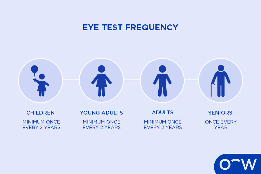
It is recommended to have an eye test at least once every 1-2 years if you do not already have existing eye conditions. If you are 50 years or older, the recommendation is once every year unless advised otherwise by your optometrist. A checkup eye test is a vital way in taking care of your eyes and vision and making sure an eye care professional can detect any possible signs of a more complex condition.
What are the Benefits of a Retinal Exam?
The benefits of a retinal exam are finding any minor changes to your retina in a timely manner to prevent further complications. It can be helpful to identify signs of an eye condition or disease early so the optometrist can refer you to an ophthalmologist for further evaluation if necessary. This can then help you start your required treatment plan early to reduce the risk of vision loss or prevent other issues from occurring.
What are the Signs that You Need a Retinal Examination?
The signs that you need a retinal examination may include symptoms such as reduced vision, a dark curtain-like shadow in your vision, double vision, excessive tearing, an eye injury, eye pain or redness. However, it is important to note that these signs are general indicators that you should see an optometrist for a standard eye test, which typically includes a retinal examination.
How Long Does the Retinal Examination Take?
A retinal examination may be as brief as regular eye tests, within which a retinal exam is conducted and these take around 30 minutes. However, this may differ depending on your condition and if the optometrist may need to run additional tests. At Oscar Wylee, our comprehensive eye tests typically take 20-30 minutes.
How Long Does it Take to Get the Results of a Retinal Exam?
It may take around 20-30 minutes for you to get the results from your eye test, which will include the findings from your retinal exam. It is important to note that a retinal exam is not separate from a complete eye test, therefore, you can receive your retinal exam results at the very end of the eye test.
What are the Eye Problems That May be Found During a Retinal Exam?
The eye problems that may be found during a retinal exam may include glaucoma, diabetic retinopathy, retinal detachment, hypertensive retinopathy and age-related macular degeneration. The eye problems found during a retinal exam are listed below.
- Glaucoma: Glaucoma refers to a group of eye problems that can cause permanent vision loss, caused by damage to the optic nerve.
- Diabetic Retinopathy: Diabetic retinopathy is an eye condition and potential complication of diabetes that can be diagnosed during an eye exam.
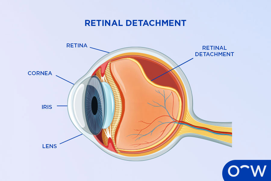
- Retinal Detachment: Retinal detachment is considered a medical emergency as the retina in the eye has pulled away from the layer of blood vessels that supply it with oxygen.
- Hypertensive Retinopathy: Hypertensive retinopathy is an eye disease that occurs when high blood pressure impairs the retina.
- Age-Related Macular Degeneration: Age-related macular degeneration causes a painless loss of central vision, due to damage to the macula which is the part of the eye that helps us with sharp vision and to see fine details.
1. Glaucoma
Glaucoma is a common eye problem that can lead to vision loss. Glaucoma may be caused by increased ocular pressure, genetics, diabetes, increased age, migraines, high-blood pressure and many other conditions. The types of glaucoma include open-angle, and closed-angle and within these types are different forms including primary glaucomas and secondary glaucomas. Acute angle-closure glaucoma is a medical emergency which requires urgent medical attention.
2. Diabetic Retinopathy
Diabetic retinopathy refers to an eye condition that can occur as a complication of diabetes and may be diagnosed during an eye exam. Diabetic retinopathy occurs when the blood vessels in the retina are damaged due to diabetes. The retina is the light-sensitive layer at the back of the eye that processes light into images. Diabetic retinopathy may not have any symptoms in its early stages, which is why patients with diabetes are recommended to get a regular eye test as an optometrist may be able to detect the issue early on. Diabetic retinopathy may be treated with laser treatment or surgery in severe cases.
3. Retinal Detachment
Retinal detachment is when the retina in the eye detaches from the layer of blood vessels that provide it with oxygen and nutrients also known as the choroid. Retinal detachment is a medical emergency and requires urgent care as the retina cannot function. Causes of retinal detachment can include ageing, an eye injury and diabetic retinopathy. Symptoms of retinal detachment can include light flashes, floaters, blurry vision and is typically painless. Treating retinal detachment typically requires surgery performed by an ophthalmologist according to Better Health.
4. Hypertensive Retinopathy
Hypertensive retinopathy is defined as an eye disease that occurs when high blood pressure damages the retina. Hypertensive retinopathy hinders blood flow to the retina, which can cause vision loss and other complications. Hypertensive retinopathy can indicate that you may have poor blood flow in other regions of your body as high blood pressure can typically affect arteries throughout the body. The symptoms of hypertensive retinopathy may not present any symptoms for a while, unless in more severe cases where low vision can occur. The risk factors for this condition can include smoking, history of cardiovascular disease, high cholesterol, obesity and high blood sugar. Treatment may include managing blood pressure levels to ensure they are reduced by keeping a healthy weight, eating balanced meals, exercising regularly and limiting alcohol consumption.
5. Age-Related Macular Degeneration
Age-related macular degeneration is characterised as a disease that develops in the macula, affecting your central vision. The two types of age-related macular degeneration are dry, which is the most common form and takes place when the macula thins out as you get older. The other type is wet (AMD), which develops when the macula is damaged by abnormal blood vessels in the back of the eye. It is considered less common and can lead to faster loss of vision. There is currently no treatment for age-related macular degeneration, therefore, an ophthalmologist will need to keep monitoring your condition through frequent eye tests.
What are the Different Eye Doctors Who Perform Retinal Exams?
The different eye care professionals who perform retinal exams are an optometrist and an ophthalmologist. An optometrist or ophthalmologist can perform eye tests which can include a retinal exam. While an optometrist is not an eye doctor, they should be your first port of call for any eye problems and can refer you to an ophthalmologist if they do identify any signs of a more complex issue. An ophthalmologist is defined as an eye doctor and eye care specialist who is qualified to manage and perform surgery on eye diseases and conditions.
Does Oscar Wylee Offer a Retinal Exam?
Yes, Oscar Wylee offers comprehensive eye exams which can include retinal exams. All stores offer retinal examinations as part of the eye test process, however, the availability of retinal examination equipment may vary between stores. You can call your local Oscar Wylee store before visiting if you require more information.
What Oscar Wylee Optometrists are Near Me?
To find an Oscar Wylee optometrist clinic near you, use our locations page or search online using the keywords, Oscar Wylee optometrist near me. The optometrist clinics in your area should easily appear in your search results if you have location settings activated on your smartphone.
Read Retinal Exam: Importance, How it Works, and Benefits in other Oscar Wylee regions and their languages.
Australia