 1300 164 990
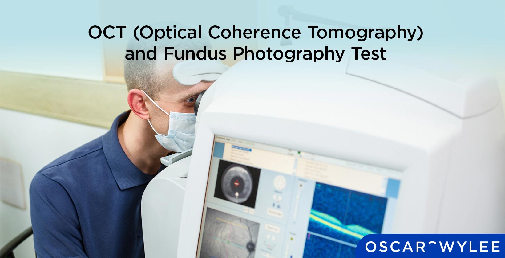
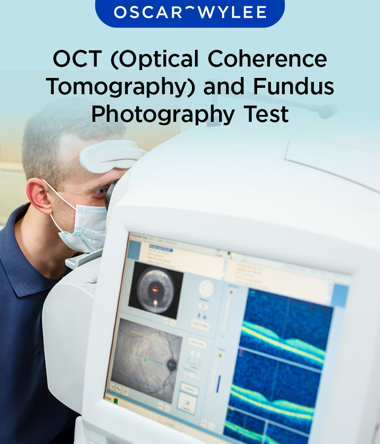
Optical Coherence Tomography (OCT) and Fundus Photography
Published on May 13th, 2024
 Australia

Optical coherence tomography (OCT) and fundus photography are both non-invasive imaging methods that take pictures of the structures in the back of the eye, including the retina, macula and optic nerve. An OCT works by using light to illuminate the retina and assessing how much light is subsequently reflected off the retina. Fundus photography works by the use of lenses and a camera and the principle of indirect ophthalmoscope. Both OCT images and fundus photography can be useful in detecting and monitoring eye conditions such as glaucoma, diabetic retinopathy, macular holes and macular puckers. The difference between OCT and fundus photography are in their method and their capabilities. For example, where an OCT can take high-resolution three-dimensional images, fundus cameras are limited to two-dimensional images, which can limit its effectiveness in diagnosing eye conditions. Keep reading to learn more about optical coherence tomography and fundus photography.
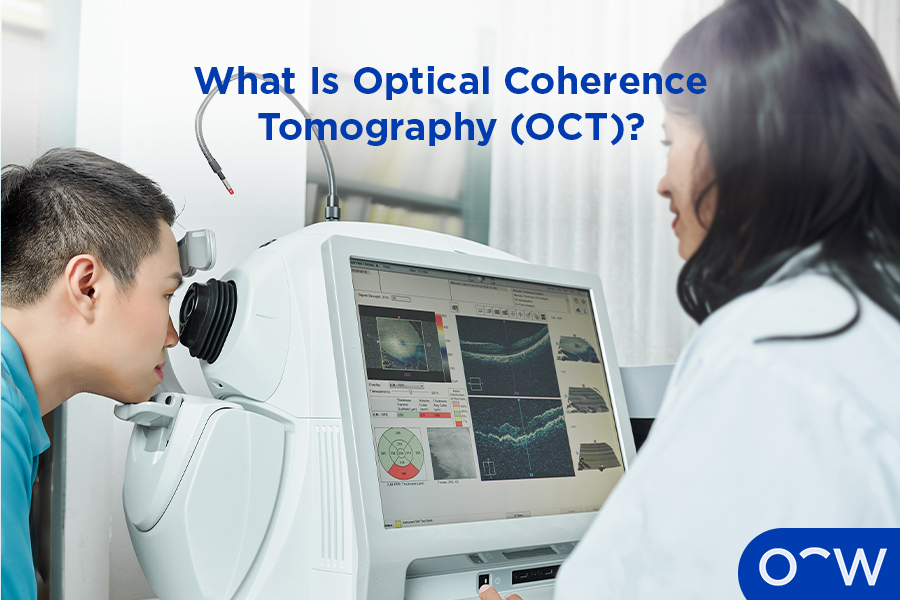
What Is Optical Coherence Tomography (OCT)?
Optical coherence tomography (OCT) is a type of non-invasive imaging that is used to create a picture of the tissues in the back of the eye, including the retina, macula and optic nerve. An OCT machine uses light waves to take an image of the retina.
How Does OCT Contribute to Eye Care?
Optical coherence tomography contributes to eye care in a major way as it is an important imaging tool that can help detect eye conditions related to the retina, macula and optic nerve, according to the American Academy of Ophthalmology. An OCT can detect eye conditions and diseases including glaucoma, diabetic retinopathy, macular holes and issues with blood vessels in the retina.
How Does Optical Coherence Tomography (OCT) Work?
Optical coherence tomography works by using near infrared light to illuminate and assess how much light is subsequently reflected off the retina and optic nerve, according to the Cleveland Clinic.
What Conditions can OCT Detect and Monitor?
An optical coherence tomography (OCT) can detect and assist in monitoring a range of eye conditions including diabetic retinopathy, glaucoma, age-related macular degeneration, macular hole and macular puckers. The eye conditions an OCT can detect and monitor are listed below.
- Diabetic retinopathy: Diabetic retinopathy is a complication of the medical condition diabetes that can change or damage the blood vessels in the retina. An OCT may help to diagnose diabetic retinopathy as it takes an image of the retina and its blood vessels.
- Glaucoma: Glaucoma refers to a group of eye diseases that cause peripheral vision loss due to optic nerve damage. An OCT takes an image of tissues and structures in the back of the eye including the optic nerve, which can help detect glaucoma.
- Age-related macular degeneration: Age-related macular degeneration is an eye condition that affects central vision and may be detected by an OCT as this type of imaging assesses the macular structure in the eye. According to Health Direct, macular degeneration is a chronic and painless eye disease that is related to aging and is caused by damage to the cells in the macula.
- Macular hole: A macular hole refers to an opening in the macula part of the retina that can lead to blurred central vision. A macular hole may be diagnosed by using an OCT that takes an image of the back of the eye.
- Macular pucker: A macular pucker, also known as epiretinal membrane, refers to a wrinkle or creases that form on the macula which can cause distorted or blurred vision. An OCT can diagnose macular pucker as it takes a detailed image of the retina, which is where the macula is found.
How is OCT Conducted?
An optical coherence tomography (OCT) is a type of non-invasive imaging method that is conducted by an optical dispenser or optometrist. The process of getting an OCT is simple and will involve a patient sitting at the OCT machine, resting their chin and forehead on the support device and keeping still while the machine takes an image. An optometrist may use eye drops to dilate a patient's pupil during an OCT, so they can see the structures of the eye more clearly, but this is not always necessary. The way an OCT is conducted is detailed below.
- Sit at the OCT machine: A patient will sit at the OCT machine, whilst an optometrist will sit behind the machine, where there is a screen on which they can see the results of the images.
- Rest chin and forehead on support device: A patient will need to rest both chin and forehead against the chin and forehead rest respectively. Both Chin and forehead rests are on the support device and both need to be resting well for good OCT imaging.
- The machine will take images: After a patient is settled, the machine will take images of the back of the eye. This should only take a few minutes and the results generally come through straight away.
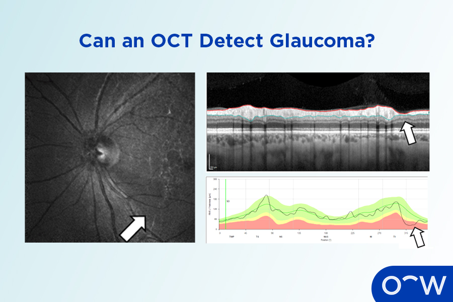
Can OCT Detect Glaucoma?
Yes, an optical coherence tomography (OCT) can detect glaucoma. Glaucoma refers to a group of eye diseases that cause peripheral vision loss due to optic nerve damage. As the optic nerve is located at the back of the eye, it can be photographed and examined during an OCT, which may help detect any issues with the structure, including glaucoma.
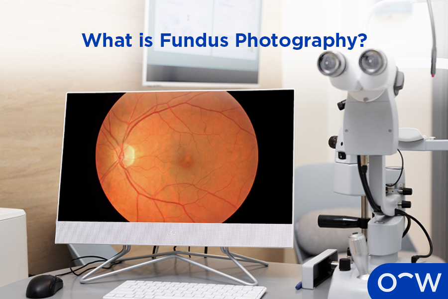
What is Fundus Photography?
Fundus photography refers to images taken of the structures in the back of the eye such as the retina, optic nerve and macula, by a fundus camera. According to Mishra and Tripathy in the paper Fundus Camera, fundus photography works by using multiple lenses and a camera and the principle of indirect ophthalmoscopy.
Why is Fundus Photography Important to Eye Health?
Fundus photography is important to eye health as it can help in diagnosing and monitoring eye conditions such as diabetic retinopathy and glaucoma. Fundus photography is a non-invasive imaging technique that captures pictures of the structures in the back of the eye.
What are the Benefits of Fundus Photography?
There are several benefits of fundus photography including that it is a type of imaging method that is non-invasive, is a quick procedure and can be used without pupil dilation. Some of the benefits of fundus photography are listed below.
- Non-invasive: Fundus photography is non-invasive, meaning that it does not require any instrument to be put into the body in any way.
- Quick: Fundus photography is a quick procedure, only taking a few minutes with results instantly visible for optometrists or eye care professionals.
- Can be used without pupil dilation: A benefit of fundus photography is that it can be used without the need to dilate a patient's pupils.
What are the Limitations of Fundus Photography?
There are several limitations to fundus photography including two-dimensional imaging, certain eye conditions affecting the clarity of images and certain fundus machines can be expensive. The limitations of fundus photography are listed below.
- Two-dimensional imaging: A fundus camera can only take two-dimensional images, as opposed to the three-dimensional capabilities of indirect binocular ophthalmoscopy, which limits how well certain conditions may be detected.
- Certain eye conditions may affect the clarity of images: Certain eye conditions, such as cataracts, in which corneal opacity is affected, may result in unclear fundus photography, according to the paper, Combining Optical Coherence Tomography and Fundus Photography to Improve Glaucoma Screening, published in the National Library of Medicine.
- Expensive: A fundus camera can be expensive to purchase and use in clinics due to recent add-ons on newer devices and the complexity and bulkiness of certain models, according to the paper, Fundus Photography in the 21st Century--A Review of Recent Technological Advances and Their Implications for Worldwide Healthcare, published in the National Library of Medicine.
What is the Process of Fundus Photography?
Fundus photography is a simple process that may differ slightly depending on the type of fundus camera used but generally involves a patient sitting still in front of a fundus camera. An optometrist or eye care professional will be on the other side of the camera and instruct it to take images of the eye. The optometrist will then assess the images and discuss the results with patients.
Is Fundus Photography Necessary?
Yes, fundus photography can be a necessary procedure as part of a routine eye test, or if an eye care professional wants to assess the structures at the back of the eye. Fundus photography allows an eye care professional to get an image of structures such as the retina, macula and optic nerve head, which can help in diagnosing eye conditions.
What Are the Key Differences Between OCT and Fundus Photography?
The key differences between OCTs and fundus photography are in how they work and their capabilities. OCT and fundus photography are similar in that they are both imaging methods that focus on capturing pictures of the back of the eye including the retina and optic nerve, however, they do not work in the same way. Optical coherence tomography works by using near infrared light to illuminate and assess how much light is reflected off the retina and optic nerve, according to the Cleveland Clinic. Fundus photography works by using multiple lenses and a camera and the principle of indirect ophthalmoscopy. There are also differences in the capabilities of these machines. OCTs are capable of taking three-dimensional, higher-resolution images of the retina, whereas fundus photography can only take two-dimensional images.
Who Needs OCT or Fundus Photography?
People who need OCT and fundus photography tests are those who are getting them done as part of their regular eye test, or those who have a specific eye condition such as diabetic retinopathy and glaucoma and need regular monitoring of these conditions. An OCT and fundus camera can be a useful tool in tracking how an eye disease is progressing.
Who Conducts an OCT or Fundus Photography?
An OCT or fundus photography can be conducted by several eye care professionals including a trained staff member, an optometrist or an ophthalmologist. An optometrist is your primary eye care provider and an ophthalmologist, sometimes called an eye doctor outside of Australia, is a medical doctor who specializes in eye care. Oftentimes an OCT will be conducted by an optical dispenser as part of pre-testing before seeing an optometrist.
How are the Results from OCT and Fundus Photography Interpreted?
The result from an OCT and fundus photography will be interpreted by an optometrist or eye care professional as soon as the images are taken. OCT and fundus photography focus on capturing images of the structures in the back of the eye such as the retina, macula and optic nerve. The results of an OCT and fundus photography may indicate if there are issues with this part of the eye and show potential eye conditions.
Read Optical Coherence Tomography (OCT) and Fundus Photography in other Oscar Wylee regions and their languages.
Australia