


 1300 164 990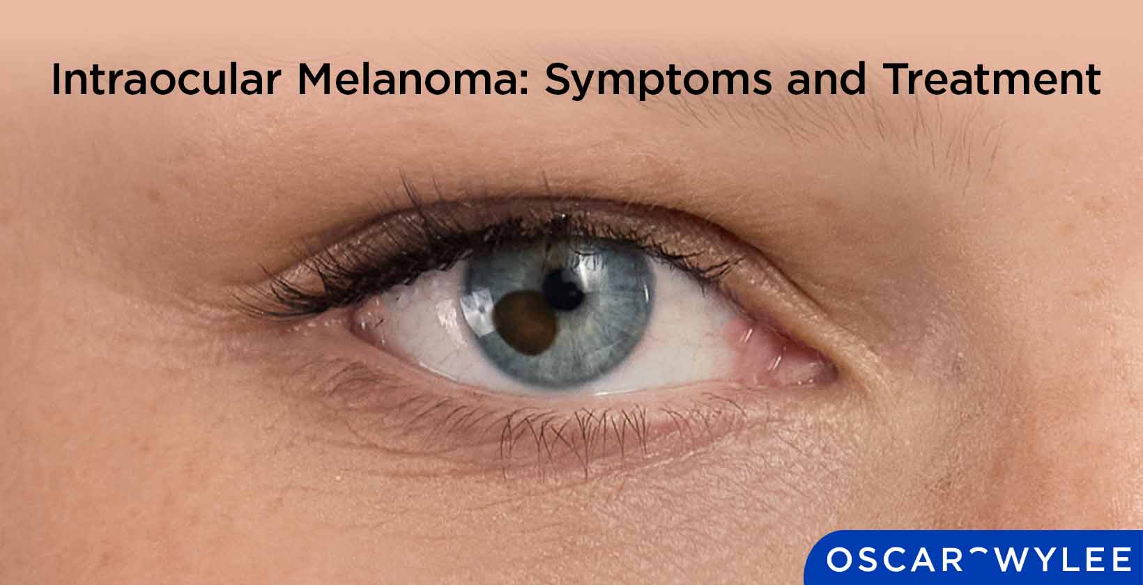

Intraocular Melanoma: Symptoms and Treatment
Published on April 10th, 2026
Intraocular melanoma is a rare form of eye cancer that develops inside the eye. It leads to the uncontrolled growth of melanocytes, which are cells that produce pigment and may cause vision changes or loss. While the term intraocular refers to the inside of the eye, melanoma can also occur on the eye's outer surface, known as conjunctival melanoma. While the exact causes of eye cancer are still unknown, genetic mutations can cause the cells to produce incorrectly and out of control. Intraocular melanoma can present symptoms such as blurred vision, flashes of light, floaters, a dark spot on the iris, changes in pupil shape, bulging of the eye and sudden vision loss. Treatment may generally include options such as surgery, radiation therapy, photocoagulation and thermotherapy.
What is Intraocular Melanoma?
Intraocular melanoma is a rare type of eye cancer that occurs inside the eye. Intraocular melanoma may originate in the middle layer of the eye, which is known as the uvea. While it is considered rare, intraocular melanoma can affect anyone, but may be more common in certain groups of people, according to the Melanoma Research Alliance. Intraocular melanoma is a serious condition, and early detection improves outcomes for patients.

How does Intraocular Melanoma Develop Inside the Eye?
Intraocular melanoma develops when pigment cells called melanocytes develop DNA mutations, causing them to grow uncontrollably. These mutated pigment cells in the eye can grow to form tumours, which lead to intraocular melanoma. This often happens within the middle layer of the eye, known as the uvea. Intraocular melanoma commonly affects vision because, as the tumour grows, it can cause blurred or distorted vision, floaters, flashes of light, and loss of peripheral vision. The location of an intraocular melanoma tumour significantly impacts symptoms, with tumours in the central macula or those that cause retinal detachment often causing immediate blurry vision or vision loss. Peripheral tumours may not cause symptoms until they grow larger.
Does Intraocular Melanoma Start From Pigment Cells in the Eye?
Yes, intraocular melanoma starts from pigment cells in the eye, which are called melanocytes. These cells produce melanin (pigment that creates eye colour), and when they develop DNA mutations, they grow uncontrollably and form tumours.
What are the Main Types of Intraocular Melanoma?
The main types of intraocular melanoma are choroidal melanoma, ciliary body melanoma, and iris melanoma. These types of intraocular melanoma are explained below.
- Choroidal melanoma: Choroidal melanoma is the most common type of intraocular melanoma in adults, and arises from the choroid layer of the eye.
- Ciliary body melanoma: Ciliary body melanoma is a rare, malignant tumour that arises from melanocytes (pigment cells) in the eye's uveal tract.
- Iris melanoma: Iris melanoma is a rare, malignant tumour that arises in the iris (coloured part of the eye).
How is Uveal Melanoma Different From Other Intraocular Tumours?
Uveal melanoma arises from the uveal tract, unlike other eye tumours such as conjunctival or cutaneous melanoma. Uveal melanoma originates from melanocytes and affects the deeper layers of the eye. It is also the most common type of intraocular cancer.
Can Intraocular Melanoma Affect the Choroid, Iris, or Ciliary Body?
Yes, intraocular melanoma can affect the choroid, iris, or ciliary body. These are parts of the uveal tract of the eye. The choroid is most commonly affected, and other areas are less frequently impacted. The location of intraocular melanoma significantly affects the symptoms a patient experiences and influences treatment choices.
What are the Symptoms of Intraocular Melanoma?
There are various symptoms of intraocular melanoma, including blurred or decreased vision, visual field defect, photopsia, flashes of light, floaters, dark spots on the iris, loss of peripheral vision, pupil shape changes, eye bulging and sudden vision loss. It is worth mentioning that many cases of intraocular melanoma are asymptomatic. A detailed list of intraocular symptoms is provided below.
- Blurred or decreased vision: Blurred or decreased vision in the affected eye is a common symptom of intraocular melanoma, with the patient struggling to see things clearly.
- Visual field defect: Visual field defect refers to areas of lost or reduced vision, blind spots (scotomas), or peripheral vision loss.
- Photopsia: Photopsia refers to a perception of flashing lights or streaks that happens without an actual source of light.
- Flashes of light: Flashes of light can include seeing flashes, streaks or wavy lines.
- Floaters (spots in vision): Floaters refer to tiny specks seen in your field of vision, according to Better Health Channel Victoria. New or increased floaters are sometimes a symptom of intraocular melanoma.
- Dark spot on the iris that enlarges: A dark spot or speck on the iris (the coloured part of the eye) that enlarges in size is a symptom of intraocular melanoma.
- Loss of peripheral vision: A significant loss of peripheral vision, which relates to an inability to see things on the side of your vision.
- Change in pupil shape: A noticeable change in pupil size (distortion) is another intraocular melanoma symptom.
- Bulging of the eye: Bulging of the eye (proptosis) is a symptom of intraocular melanoma, often accompanied by redness and pain. This is only usually seen in late-stage cases.
- Sudden vision loss: Sudden vision loss may occur in intraocular melanoma patients, but it is usually a sign of advanced tumour growth or related to retinal detachment. Sudden vision loss is a medical emergency. If you experience a rapid change in your vision, please contact your optometrist or healthcare professional immediately, or visit the nearest emergency department.

How do Early Signs of Intraocular Melanoma Appear?
Early signs of intraocular melanoma appear as subtle vision changes or spots and may be difficult to notice. Early signs of intraocular melanoma include new or increased floaters developing and blurred or decreased vision. Early detection of intraocular melanoma is difficult, and regular eye tests are important. If you notice any changes in your vision, it is important to book an eye test with an optometrist so that the health of your eyes can be assessed.
What Causes Intraocular Melanoma?
The causes of intraocular melanoma are not fully understood, but are related to mutations occurring in the pigment cells of the eye. There are various risk factors that are related to intraocular melanoma, including genetic mutations, light-coloured eyes, fair skin or sun sensitivity, older age, ocular melanocytosis, pre-existing eye nevi, BAP1 tumour predisposition syndrome, and possible occupational or environmental exposure. A detailed list of different risk factors for intraocular melanoma is provided below.
- Genetic mutations (e.g., in GNAQ, GNA11, BAP1): Genetic mutations that turn normal melanocytes into malignant tumours are a main risk factor for intraocular melanoma. 'The GNAQ and GNA11 genes are mutated in almost 80–90% of uveal melanomas in a mutually exclusive pattern', according to P. Silva-Rodriguez's research paper titled 'GNAQ and GNA11 Genes: A Comprehensive Review on Oncogenesis, Prognosis and Therapeutic Opportunities in Uveal Melanoma' published in 2022. Mutations in the BAP1 gene are also strongly associated with intraocular melanoma.
- Light-coloured eyes (blue, green, grey): Having light-coloured eyes is a significant and well-documented risk factor for intraocular melanoma, although the reason is not completely clear.
- Fair skin/sun sensitivity: There is a significant risk of intraocular melanoma developing in patients with fairer skin or who are generally more sensitive to the sun.
- Older age (especially over 50): Intraocular melanoma is more common in those over the age of 50, and is rarely found in children, according to the Melanoma Research Alliance, although the exact reason for this is not fully known. It is important to note that the incidence of intraocular melanoma is rare overall, according to Cancer Council Australia.
- Ocular or oculodermal melanocytosis: Ocular or oculodermal melanocytosis is characterised by blue or grey patches on the eye or skin. Ocular or oculodermal melanocytosis is a significant risk factor for developing intraocular melanoma.
- Pre-existing eye nevi: Pre-existing eye nevi are pre-existing benign growths called choroidal nevi (eye moles or freckles) that may develop into intraocular melanoma, specifically uveal melanoma.
- BAP1 tumour predisposition syndrome: BAP1 tumour predisposition syndrome is an inherited condition caused by a mutation in the BAP1 gene that significantly increases the risk of developing specific cancers at a younger age, including uveal melanoma. BAP1 tumour predisposition syndrome is associated with uveal melanoma, according to R. Pilarski's research paper titled 'BAP1 Tumour Predisposition Syndrome', published in 2016.
- Possible occupational/environmental exposure (e.g., welding, chemicals): Possible occupational/environmental exposure, such as welding or exposure to certain chemical solvents, has been linked to the development of intraocular melanoma. A case-controlled study conducted by E. A Holly, D. A Aston, D. K Ahn, and A. H Smith, titled 'Intraocular melanoma linked to occupations and chemical exposures', published in 1996, found an increased risk of uveal melanoma for occupational groups who had intense exposure to ultraviolet light, welding exposure, and asbestos exposure. Although this study suggested a link to occupational exposures like welding, research is ongoing to confirm that these environmental factors are linked to intraocular melanoma.
What Risk Factors are Linked to Intraocular Melanoma?
Risk factors linked to intraocular melanoma include light eye colour, genetics, and increasing age, according to Cancer Council Australia. These factors may increase susceptibility to intraocular melanoma. People with light eyes, fair skin, and hair that is blonde or red have a higher risk of developing intraocular melanoma due to a lack of melanin, which protects against harmful UVA and UVB rays. While UV is a well-known risk factor for skin melanomas and conjunctival melanomas, there is mixed evidence to suggest that UV exposure is linked to intraocular (uveal) melanomas. Genetic predisposition also plays a role; for example, people with inherited skin or mole disorders have a higher risk of developing intraocular melanoma. The risk of developing intraocular melanoma increases with age, with the average diagnosis being around 55-60 years old. Environmental exposure also contributes to the increased risk of developing the disease. Risk awareness aids in prevention, and monitoring is important.
Always seek the advice of a qualified optometrist, ophthalmologist, or other healthcare professional with any questions you may have regarding a medical condition or eye health. If you experience any changes in your vision, including blurred vision, sudden flashes of light, new floaters (spots or wavy lines), or a growing dark spot on the iris, you should consult a qualified optometrist or ophthalmologist immediately.
What are the Treatment Options for Intraocular Melanoma?
The treatment options for intraocular melanoma aim to preserve vision and the eye itself, and include radiation, laser therapies, or surgery. The most common types of these treatments are listed below.
- Plaque Brachytherapy (radiation): Plaque Brachytherapy involves a radioactive disc being temporarily stitched onto the outside of the tumour, usually for 3-7 days, which administers targeted radiation to the tumour.
- Transpupillary Thermotherapy (laser therapy): Transpupillary Thermotherapy (TTT) involves an infrared laser that heats and therefore destroys cancer cells associated with intraocular melanoma tumours. This treatment is often used in addition to radiation rather than as a standalone treatment.
- Enucleation (surgery): Enucleation refers to the removal of the entire eye, which usually only occurs for very large tumours. An iridectomy or iridocyclectomy may be performed, which is the removal of a part of the eye where the tumour is located. Local resection refers to the surgical removal of the tumour itself.
Treatment for intraocular melanoma is managed by specialist ophthalmologists and oncologists. Your optometrist plays a vital role in early detection and referral.
How Does Surgery Compare to Radiation for Intraocular Melanoma Treatment?
Surgery for intraocular melanoma removes the tumour or the eye, while radiation targets tumour cells without removal. Both of these treatments aim to control cancer; surgery is more invasive, and radiation can help preserve the structure of the eye. Radiation, specifically plaque brachytherapy, is more often used for small to medium-sized tumours, and surgery is reserved for large tumours that cannot be treated with radiation. The choice of either depends on your healthcare professional's advice, usually based mostly on the severity of the tumour. Outcomes of these treatments vary case by case.
Can Intraocular Melanoma be Treated Without Removing the Eye?
Yes, intraocular melanoma can be treated without removing the eye, most commonly when radiation and laser methods are used. These often eye-preserving treatments do exist, and it is often early detection that allows these less invasive options to be provided to a patient. Preservation of the eye aims to improve quality of life for patients, but treatment choice is case-based.
How Can Vision Be Managed After Intraocular Melanoma Treatment?
Vision is managed after intraocular melanoma treatment through corrective lenses, rehabilitation, and targeted therapies. Corrective lenses refer to prescription glasses that can improve clarity, with patients often needing a new prescription to adjust to changes in their vision following treatment. Tinted lenses and sunglasses are also recommended to manage light sensitivity, which is a common side effect of radiation therapy, specifically. Rehabilitation refers mostly to patients who experience permanent vision loss, and includes low vision aids, accessibility tools, and occupational therapy. It is important to note that vision loss after treatment is often due to the location of the tumour or side effects of radiation, rather than the surgery itself. Target therapies that can reduce complications that may follow treatment include anti-vascular endothelial growth factor (anti-VEGF) injections and laser therapy. Regular monitoring is required following intraocular melanoma treatment, including follow-up care to track recovery. Although the management options mentioned above can support daily function, long-term care is needed to support full recovery and optimal eye health.
Intraocular melanoma is a complex condition, and every patient's recovery journey is unique. It is essential that you consult your ophthalmologist or eye care professional before starting any new treatment or rehabilitation program. Only a qualified specialist can provide a tailored management plan based on your specific clinical history and response to therapy.
What Types of Glasses Help After Intraocular Melanoma Treatment?
Many different types of glasses can help patients after intraocular melanoma treatment, including UV-protection glasses, wrap-around glasses, polarised lenses, tinted lenses, transition lenses, prescription glasses, protective glasses, and anti-reflective coated lenses. A detailed list of different types of glasses that can help patients after intraocular melanoma treatment is provided below.
- UV-protection sunglasses (UV400 or 100% UVA/UVB blocking): UV-protection sunglasses, specifically UV400 or 100% UVA/UVB blocking sunglasses, are highly recommended after intraocular melanoma treatment because they protect the eyes from further damage and increased light sensitivity that may occur after procedures like radiation.
- Wrap-around sunglasses: Wrap-around sunglasses are recommended after intraocular melanoma treatment because they provide full side protection and protect the eyes from harmful UVA and UVB rays, which may trigger further damage.
- Polarised lenses: Polarised lenses may help reduce glare and improve comfort for patients experiencing light sensitivity post-treatment, as the eyes are often highly sensitive to light following this, and polarised lenses reduce glare and improve visual comfort.
- Tinted lenses: Tinted lenses are beneficial for patients who have recently undergone treatment for intraocular melanoma, particularly medium-dark to dark tints, or brown, amber, and grey tints. Medium-dark to dark tints in sunglasses with a lens category of 3 or 4 provide maximum protection against harmful UVA and UVB rays. Brown and amber tints can help with enhancing contrast and improving depth perception, and grey tints help provide a neutral colour perception.
- Photochromic (transition) lenses: Photochromic, or transition lenses, may be highly beneficial for patients after intraocular melanoma treatment, as they can improve light sensitivity symptoms associated with recovery. They are ideal for recovery because they automatically become clear indoors while also protecting eyes from harmful UVA and UVB rays outdoors. They can also improve eye strain and eye fatigue symptoms.
- Prescription glasses: Prescription glasses may be necessary for patients if intraocular melanoma treatment causes refractive changes such as new myopia, hyperopia, or astigmatism.
- Protective glasses/shields (during early healing period): Protective glasses or shields are crucial in protecting the eyes from physical injury after intraocular melanoma treatment.
- Anti-reflective (AR) coated lenses: Anti-reflective (AR) coated lenses are recommended to patients after undergoing intraocular melanoma treatment to manage side effects such as light sensitivity, seeing halos, and eye fatigue.
While the eyewear listed above is particularly beneficial for those recovering from intraocular melanoma treatment, UV-protective glasses are strongly recommended for everyone, regardless of medical history. Consistent use of high-quality UV400 or 100% UVA/UVB blocking lenses is an important step in maintaining long-term eye health and protecting against sun-related damage for all individuals.
Can Protective or Prescription Glasses Improve Vision After Treatment?
Yes, protective or prescription glasses may improve vision after intraocular melanoma treatment. Protective or prescription glasses (if there is a refractive error present) may help to optimise remaining visual function and comfort following intraocular melanoma treatment, by enhancing clarity and reducing eye strain. Patients may require a new prescription after undergoing intraocular melanoma treatment to adjust to changes in vision. Tinted lenses and sunglasses are recommended for patients post-treatment, as they can help with light sensitivity symptoms that are common after radiation treatment. UV-protection sunglasses are recommended as they help reduce glare and protect the eyes from external environmental stressors. Protective or prescription glasses help support rehabilitation after surgery and improve overall comfort for patients. It is important to note that the visual prognosis after treating intraocular melanoma is highly variable.
How Can Light Sensitivity Be Reduced in Intraocular Melanoma Patients?
Light sensitivity may be reduced in intraocular melanoma patients by using UV protective eyewear and lenses, environmental adjustments, and medical interventions. Protective eyewear and lenses include polarised sunglasses, wraparound sunglasses, specialised tints, and photochromic lenses to reduce glare and protect the eyes from harmful UVA and UVB rays. Blue light filters can help reduce eye strain caused by digital screens and artificial lights. Environmental adjustments include limiting bright exposure and using control lighting where possible, such as dimmer switches. Patients may also find it helpful to avoid fluorescent lights and use diffusers to spread light to make it less intense. Medical interventions involve the use of lubricating eye drops to maintain moisture in the eyes and lessen the effects of light sensitivity. Symptom relief for intraocular melanoma patients is important and supports daily function and well-being.

What Role do UV Protective Sunglasses Play for Intraocular Melanoma Patients?
UV protective sunglasses shield eyes from harmful radiation and may reduce irritation and risk for intraocular melanoma patients. UV protective sunglasses can help prevent further damage to the eyes caused by harmful UVA and UVB rays, as evidence suggests that UV radiation can contribute to DNA damage in the iris. For patients who have undergone intraocular melanoma treatments that can damage the surface of the eye, UV-protective glasses can help prevent secondary eye conditions such as cataracts. More generally, UV-protective sunglasses are crucial in protecting the eyes and the delicate skin around the eyes from developing skin cancer. The eyes may be more sensitive to the sun after intraocular melanoma treatment, making UV-protection important to ease light sensitivity.
What Vision Problems Can Intraocular Melanoma Cause?
Intraocular melanoma can cause various vision problems, including blurred vision, loss of peripheral vision, partial or complete loss in one eye, seeing flashes of light, floaters, distorted vision, dark spots, reduced visual acuity, double vision, pupil size changes and increased light sensitivity. A detailed list of vision problems caused by intraocular melanoma is provided below.
- Blurred vision: Blurred vision is a common vision problem caused by intraocular melanoma. It involves poor vision in the affected eye, which can be blurry or hazy.
- Loss of peripheral (side) vision: Loss of peripheral vision describes loss of side vision, which can make it difficult to see things on the side.
- Partial or complete vision loss in one eye: Partial or complete vision loss in one eye is a serious vision problem caused by a severe case of intraocular melanoma.
- Seeing flashes of light (photopsia): Seeing flashes of light, also known as photopsia, is a recognized vision problem associated with intraocular melanoma. It is often described as lightning streaks or shooting stars in the field of vision.
- Floaters (spots or shapes drifting in vision): Floaters are tiny specks, spots, or wavy lines that are a common vision problem associated with intraocular melanoma.
- Distorted vision (straight lines appear wavy): Distorted vision where straight lines appear wavy or curved is a vision problem linked to intraocular melanoma.
- Dark spots or shadows in the visual field: Dark spots or shadows in the visual field, including floaters and other visual disturbances, are a common vision problem caused by intraocular melanoma.
- Reduced visual acuity: Reduced visual acuity relates similarly to blurred or generally poor vision, often caused by intraocular melanoma. Objects may appear blurry, hazy, or out of focus.
- Double vision (less common): Double vision, or diplopia, is a less common vision problem caused by intraocular melanoma, where the patient may experience the perception of two images of a single object, either horizontally, vertically, or diagonally.
- Changes in pupil shape or size: Changes in pupil shape or size are a common visual side effect that can affect intraocular melanoma patients. The pupil may become larger as the cancer affects the iris, or it may become distorted, ragged, or asymmetrical.
- Increased light sensitivity (photophobia): Increased light sensitivity, or photophobia, is a less common symptom than flashing lights, but it may occur particularly if the tumour causes inflammation or irritation.
How does Intraocular Melanoma Tumour Growth Affect Vision and Eye Function?
Intraocular melanoma tumour growth disrupts normal eye structures and vision signals as the tumour can grow and impact the retina, lens, or ciliary body. Intraocular melanoma tumour growth can cause vision impairment, including blurred or distorted vision, loss of peripheral vision, flashes and floaters, and in some severe cases, complete loss of vision. The disease can affect eye function in some cases, causing secondary problems such as retinal detachment, changes in pupil size, secondary glaucoma, visible dark spots, and bulging eye (proptosis), which is only usually seen in late-stage cases. Intraocular melanoma may interfere with light processing, and retinal function is often affected. Patients' eye pressure may change, and their function can decline over time.
It is important to note that Oscar Wylee offers optometry services only; this article is for educational purposes only and is not a substitute for professional medical advice, diagnosis, or treatment.