 1300 164 990
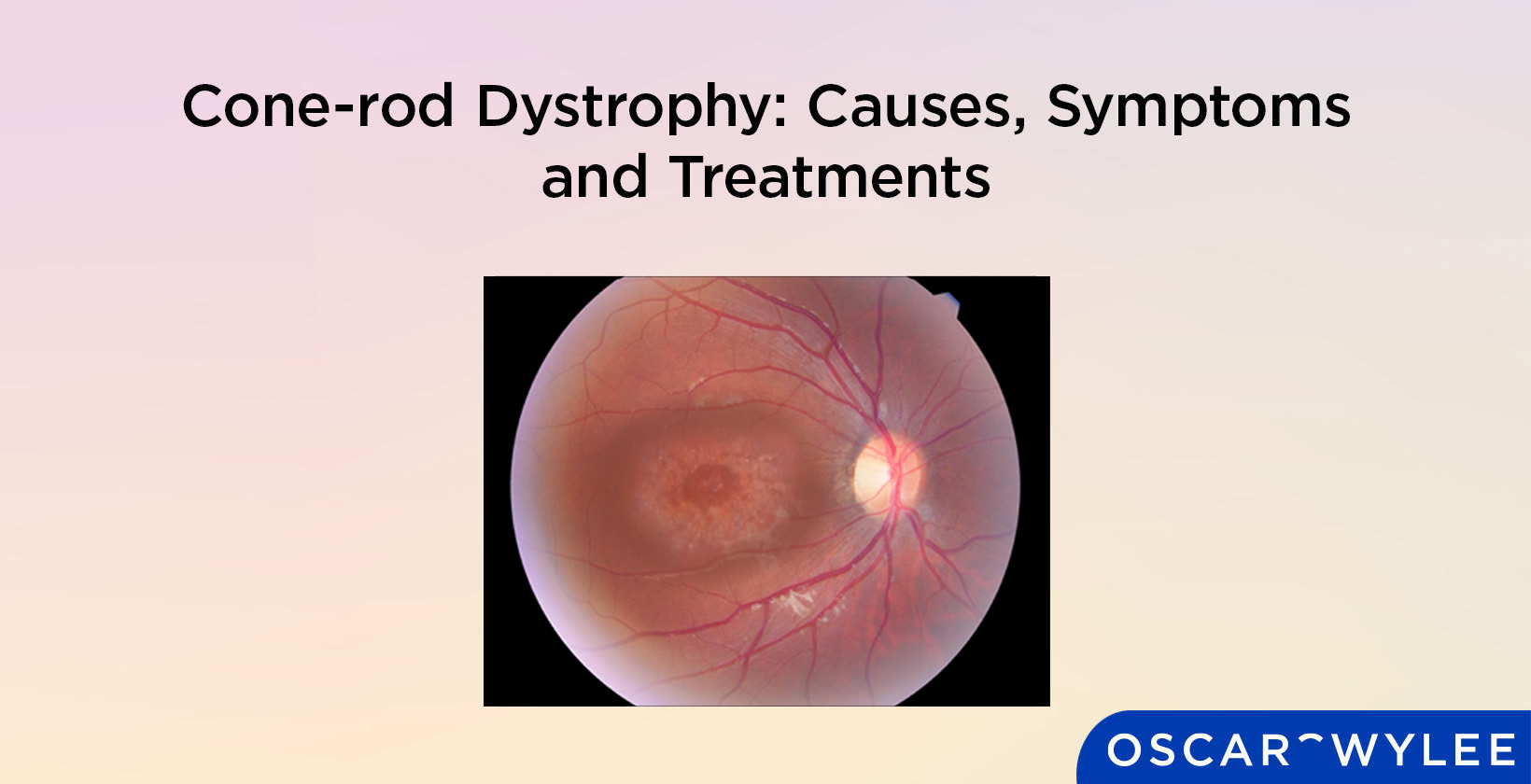

Cone-rod Dystrophy: Causes, Symptoms and Treatments
Published on July 5th, 2024
Cone-rod dystrophy refers to an eye condition that affects light-sensitive cells in the retina called cones and rods. The cones and rods in the eye help detect light and turn this light into electrical signals that will be sent to the brain to help a person see. If a person has cone-rod dystrophy, these light-sensitive cells, also called photoreceptors, will begin to deteriorate. The symptoms of cone-rod dystrophy can include decreased visual acuity, light sensitivity, poor vision at night or in low levels of light and issues with colour vision. There is no cure for cone-rod dystrophy, with treatment for this condition revolving around managing a patient's symptoms such as decreased visual acuity or light sensitivity. Keep reading to learn more about cone-rod dystrophy.
What is Cone-rod Dystrophy?
Cone-rod dystrophy (CORD) is an inherited eye disorder that affects the light-sensitive cells, or photoreceptors, in the retina called rods and cones. The retina is the light-sensitive layer at the back of the eye. Rods and cones are the cells in the retina that detect light and turn this light into electrical signals that are sent to the brain to help us see. Cone cells are responsible for color vision and rod cells are responsible for vision in low light. According to the Genetic and Rare Diseases Information Centre (GARD), cone-rod dystrophy causes the rods and cones to deteriorate, which leads to vision loss. The cones will be the first photoreceptor cells to deteriorate with cone-rod dystrophy. With the similar eye condition rod-cone dystrophy, the rods are the first photoreceptor cells to deteriorate.

How does Cone-rod Dystrophy Affect a Person's Vision?
Cone-rod dystrophy typically affects a person’s colour vision, visual acuity and sensitivity to light. According to Eye Research Australia, the age at which vision is affected and the rate of progression for cone-rod dystrophy varies for different people.
What is the Cause of Cone-rod Dystrophy?
Cone-rod dystrophy is typically caused by gene mutations. Certain genes help the photoreceptors function and provide their structure, according to MedlinePlus. Changes or mistakes in these genes can lead to the photoreceptors in the retina not working correctly, according to Eye Research Australia. The cone cells are generally the first photoreceptor that deteriorates in cone-rod dystrophy, whereas the rods will deteriorate first if someone has rod-cone dystrophy.
What are the Symptoms of Cone-rod Dystrophy?
The main symptoms of cone-rod dystrophy are decreased visual acuity, light sensitivity, poor vision at night or in low levels of light and issues with colour vision. According to the Genetic and Rare Diseases Information Centre (GARD), the symptoms of cone-rod dystrophy can start to appear at varying ages, including from childhood to adulthood. The symptoms experienced, and the severity of these symptoms at the age at which they begin can vary for different people.
What are Standard Procedures in Diagnosing Cone-rod Dystrophy?
The standard procedures that are carried out in the diagnosis of cone-rod dystrophy include eye tests such as a visual acuity test, a colour vision test, an electroretinogram and the consideration of a patient's symptoms and family history. Cone-rod dystrophy will be diagnosed by an eye care professional, such as an optometrist or ophthalmologist. According to the National Organisation for Rare Disorders, an eye care professional will consider a patient's symptoms and family before moving on to ophthalmological exams such as a peripheral vision test, a visual acuity test, a colour vision test and an electroretinogram. The standard procedures for diagnosing cone-rod dystrophy are listed below.
- Consider symptoms: Considering a patient's symptoms and family history is the first step in diagnosing cone-rod dystrophy. Symptoms that may indicate a person has cone-rod dystrophy include light sensitivity, reduced visual acuity and issues seeing colours correctly.
- Consider family history: Considering a patient's family history in regard to eye conditions and cone-rod dystrophy is an important step in diagnosing this eye condition as it is generally inherited.
- Visual acuity test: A visual acuity test measures how sharp a person’s vision is and helps in the diagnosis of cone-rod dystrophy. A visual acuity test is typically conducted by an optometrist, who has a patient read from a letter chart. This letter chart, often a Snellen chart or LogMAR chart, has letters descending in size and will be 6 metres or 20 feet away from the patient. A patient will read each line of letters until they can no longer.
- Colour vision test: A colour vision test can help diagnose cone-rod dystrophy as one of the main symptoms of this condition is issues with colour vision. A colour vision test is typically either an Ishihara test (colour plate test) or a colour hue test ( colour arrangement test).
- Electroretinogram: An electroretinogram is a test that measures the electrical response of the rods and cones.
What are the Treatment Options for Cone-rod Dystrophy?
As there is no cure for cone-rod dystrophy, treatment focuses on relieving the symptoms of this condition as it presents in each individual. This could mean wearing sunglasses to help with light sensitivity, glasses that help with colour vision issues, or visual aids to help with reduced visual acuity.
What are the Daily Challenges of Living with Cone-rod Dystrophy?
The daily challenges of living with cone-rod dystrophy will be dependent upon the individual and how progressed the condition is. In the earlier stages of cone-rod dystrophy, symptoms such as light sensitivity, reduced visual acuity or issues with colour blindness, may not have progressed enough to impact daily life. In later stages, additional symptoms such as loss of peripheral vision and nystagmus, as well as decreasing visual acuity, may make it difficult to move autonomously, according to Hamel CP. in the paper Cone rod dystrophies. Decreased visual acuity may make tasks such as driving or reading more difficult. Increased light sensitivity may also make it hard for people to be outside without sunglasses.
What Role Does Genetics Play in Cone-rod Dystrophy?
Genetics play a major role in cone-rod dystrophy, as this condition is caused by an inherited gene mutation. According to MedlinePlus, mutations in over 30 genes are associated with causing cone-rod dystrophy. According to Skorczyk-Werner, A., Chiang, WC., Wawrocka, A. et al., genes that cause cone-rod dystrophy can be inherited in autosomal dominant, autosomal recessive or X-linked recessive ways.
How does Cone-rod Dystrophy Connect to Color Vision Deficiency?
The connection between cone-rod dystrophy and colour vision deficiency is the breakdown of the cone cells. The cone cells are light-sensitive cells in the retina that are responsible for colour vision. Cone-rod dystrophy is an eye condition that causes a breakdown of the cone cells. When this occurs, colour vision can be affected.
Can Cone-rod Dystrophy be Cured?
No, cone-rod dystrophy can not be cured. According to the National Organisation for Rare Disorders, treatment for cone-rod dystrophy focuses on managing symptoms for the individual, as opposed to trying to cure the condition.
Is Cone-rod Dystrophy Painful?
Cone-rod dystrophy will generally only cause discomfort or pain if a person has increased light sensitivity due to this condition. Light sensitivity, also known as photophobia, is a condition in which the eyes are susceptible to light. For people with light sensitivity, being in bright light may cause discomfort or pain in the eyes.
What is the Difference Between Cone-rod Dystrophy and Corneal Dystrophy?
The main difference between cone-rod dystrophy and corneal dystrophy revolves around which parts of the eye they affect. Cone-rod dystrophy refers to an eye condition in which the light-sensitive cells deteriorate. Corneal dystrophy refers to an eye condition in which abnormal material accumulates in the clear part of the eye, or the cornea, according to the National Organisation for Rare Disorders. These conditions are similar in that they are both genetic conditions, inherited from family members.
What is the Difference Between Cone-rod Dystrophy and Fuchs Dystrophy?
There are many differences between cone-rod dystrophy and Fuchs dystrophy, with the main two being the part of the eye they affect and how this part is affected. Fuchs dystrophy is a type of corneal dystrophy. Corneal dystrophies are a group of eye conditions that involve changes to the cornea. Cone-rod dystrophy refers to an eye condition that affects the photoreceptors, or the light-sensitive cells, in the eye. These conditions are similar in that they can both be inherited from family members.