 1300 164 990
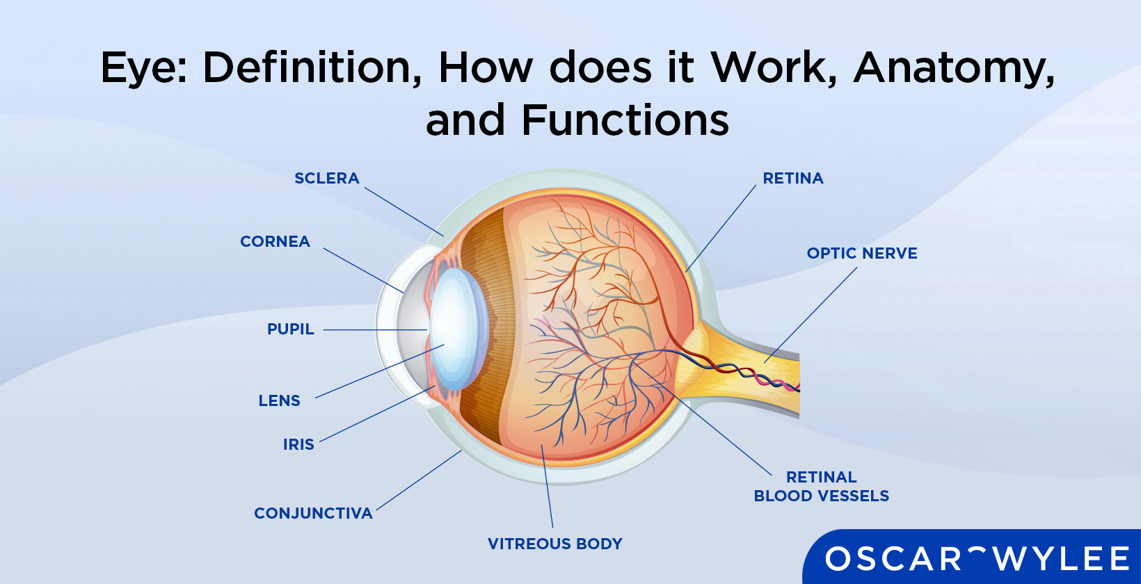

Eye: Definition, How does it Work, Anatomy, and Functions
Published on February 19th, 2023
Updated on November 15th, 2024
 Australia
The eye is defined as the part of the human body that creates and controls how and what a person is capable of seeing. The anatomy of the eye is made up of several parts, each with its own purpose in protecting the eye or assisting with sight. The eye receives light through the pupil which is sent to the lens which bends and transforms shapes in order to send light to the retina and macula, which creates images to send to the brain through the optic nerve. The parts of the eye include the outer layer, which receives light and protects the other layers, the middle layer, which nourishes the eye and contains the most blood of the eye, and the inner layer, which receives light and transforms the light into images to send to the brain.
Eye colour and eye shape vary heavily from person to person. The eye-seeing ability is affected by damage, diseases and eye conditions, and blue light wavelengths. Other animals, such as eagles, have different makeup in their eye anatomy that affects their vision and allows them to see clearer or sharper than the human eye is capable of seeing.
What is the Eye?
The eye is an organ in the human body that controls sight and allows a person to see. It is an asymmetrical globe located in the head. The iris of the eye can be different colours, or pigmentations. The eye is similar to an organic camera located in the head's socket, it is capable of focusing on one single object for the sake of visual clarity and perceiving as much detail as possible while blurring any irrelevant or non-focused aspects of the immediate image.
The evolution of the eye is not a clear matter. The eye was believed to have evolved from organic mechanics that helped the body to find and keep a circadian rhythm: the ability to tell night and day outside for the sake of knowing when to rest and when to wake up and take action in the daytime, according to I R Schwab, from the University of California’s Department of Ophthalmology & Vision Science, during a lecture on the development of the ocular toolkit.
What is the white part of the eye called?
The white part of the eye is called the sclera. The sclera shields the eye from injury and helps the eye to stay in a globe shape.
How Does the Human Eye Work?
The human eye works by receiving light through the pupil, focusing light through the lens onto the macula and retina. The eye creates sight by absorbing light wavelengths from the outside world through the outer layer of the eye. The outer layer of the eye contains the white part of the eye, called the sclera, the cornea and the iris. The eye takes this light and uses the inner layer of the eye to create an image from the light the outer layer received. The inner layer of the eye contains the retina and the optic nerve. The middle layer of the eye is used to nourish the eye and contains the choroid and the iris.
The eye must stay moist to function properly, and has the 3 major layers of the tear film to create tears which contain oils that supply constant lubrication and nourishment.
What are the Different Parts of an Eye?
There are 19 main part of the eye. The components of the eye and how they function are listed below.
- The choroid: The choroid is located between the sclera and the retina and is where 85% of the eyes’ blood is stored.
- The ciliary body: The ciliary body is one of three parts that forms the middle layer of the eye and provides nutrients for the lens and cornea.
- The cone cells: The cone cells are photoreceptors, a structure that helps convert light into signals that are then sent to the brain. Cone cells control colour vision.
- The cornea: The cornea is the transparent outer layer at the front of the eye. The cornea helps the eye focus on light.
- The conjunctiva: The conjunctiva is the membrane that covers the eyelids and the surface of the eye. The conjunctiva protects the eye and keeps it wet.
- The crystalline lens: The crystalline lens or more commonly known as the lens, is a transparent structure that helps the eye to focus light onto the retina.
- The fovea: The fovea is a small depression located at the centre of the retina that is responsible for direct and central vision in the eye.
- The iris: The iris is the visible, coloured part of the eye. It controls the amount of light that enters the eye.
- The Lens: A different term for the crystalline lens.
- The macula: The macula is the centre of the retina, located at the back of the eye. It converts light seen in the eye into images.
- The optic disc: The optic disc is an opening that communicates signals between the optic nerve and photoreceivers, allowing us to see.
- The optic nerve: The optic nerve controls and transmits all visual information to the brain.
- The pupil: The pupil is in the iris. The pupil is responsible for the volume of light that enters the eye.
- The retina: The retina is a structure at the back of the eyeball. The retina converts the light that enters the eye into images that are sent to the brain.
- The rod cells: The rod cells are photoreceptors that are sensitive to low levels of light. They will help you see at nighttime.
- The sclera: The sclera is the visible white of the eyeball. The sclera shields the eye from injury and helps keep its shape.
- The tear film: The tear film refers to several layers of fluid that cover the surface of the eye. The tear film keeps the eyes lubricated and stops them from going dry.
- The vitreous body: The vitreous body is a gel substance found throughout the eye. The vitreous body keeps the eye in its round shape.
- The zonules: The zonules are tiny fibres found in the eye. The zonules help the lens in place.

1. Choroid
The choroid is a layer of the eyes located between the sclera and the retina. According to ScienceDirect, the choroid is made up predominantly of blood vessels and accounts for much of the blood flow found within the eyes. The choroid’s first function in the eye is to use the blood flow to provide nutrients and sustenance to other vital parts of the eye, such as the retina, macular and optic nerve. The second function of the choroid is to shield the eye from possibly harmful light and reflection. The protective part of the choroid is what causes ‘red eye’ in photography.
If the choroid is damaged the retina begins bleeding. The damage eventually results in the loss of peripheral vision and central vision.
2. Ciliary Body
The ciliary body is a circular ring that is connected to the iris. The ciliary body is one of three parts that form the middle layer of the eye. The other parts of the middle layer are the choroid and the iris. The first function of the ciliary body is to hold the lens in place in the eye using the ciliary muscle which also helps to accommodate the lens, making the lens able to change shape and focus the eyes. The second function of the ciliary body is to use the ciliary processes to produce a fluid in the eye called aqueous humour. Aqueous humour is a visible, clear fluid that is vital for nourishing the eye and helps the eye to maintain its shape.
Damage to the ciliary body results in blurred vision and an inability to focus. Damage to the ciliary body results in complete vision loss if it is left untreated.
3. Cone Cells
Cone cells are found near the macula of the eye and help to perceive colours and details. There are 6 million cone cells found within the eye. The first function of cone cells is to help convert light into signals to send to the brain as they are photoreceptors which are cells in the eye that are sensitive to light. Cone cells are one of two types of photoreceptors found within the eye. The other type of photoreceptor is the eye rods and their variations. There are three types of cone cells: red-sensing cone cells, blue-sensing cone cells and green-sensing cone cells. Each type of cone cell functions to help the eye perceive the pigmentation of the cone cells sensing colour.
Cone cells are responsible for colour vision within the eye. The second function of cone cells and rod cells is to translate light into electric signals and then send them to the brain through the optic nerve.
Damage to the cone cells results in sensitivity to light, less visual information when looking straight ahead and difficulty seeing colour.
4. Cornea
The cornea is the clear outer layer at the front of the eye, according to the National Eye Institute. It is located in the very front portion of the eye, directly covering the pupil, iris and anterior chamber. The first function of the cornea is to control how much light enters the eyes and focus the light that is received. The cornea bends and refracts any light the eye receives by changing size, becoming smaller or larger, depending on the volume of light entering the eye. The cornea is the first contact light has with the human eye and serves to focus the majority of the light the eye receives.
The cornea’s second function is to protect the eye by filtering out harmful glare from the eye, such as ultraviolet rays from sunlight. The cornea is equipped to deal with minor damage, such as cuts, scars or other abrasions.
Heavy damage to the cornea results in distorted vision and a lesser ability to see light.
5. Conjunctiva
The conjunctiva is a thin layer of tissue, referred to as a mucous membrane, that covers the whites of the eye and lines the inside of the eyelid. The conjunctiva’s layer of tissue is clear and see-through. It is located in the outer layer of the eye. The first function of the conjunctiva is to secrete fluids that keep the eyes lubricated and protected from infections and outside bacteria and bodies, such as dust or other bodies.
The conjunctiva itself is made up of three aspects, the bulbar conjunctiva, the palpebral conjunctiva and the fornix conjunctiva. The bulbar conjunctiva is the section that covers the sclera but not the cornea. The palpebral conjunctiva is the section that covers the eyelid in both its upper and lower parts. The fornix conjunctiva is located between the other two sections, the fornix conjunctiva allows the eyelid and eyeball freedom of movement. The second function of the conjunctiva is to ensure no foreign body or object slips behind the eye, such as contact lenses.
Damage to the conjunctiva affects the eyes' appearance, making them discoloured and visibly injured and causing conditions such as dry eye and blurred vision.
6. Crystalline Lens
The crystalline lens is a natural focusing lens in the eyes that provides a portion of the optical power found within the eye. It is also simply referred to as the lens of the eye. The first function of the lens is for bending and focusing light by changing shape with its natural elasticity. The lens uses its changing shape to shine the light it receives directly into the retina, which begins the process of changing outside light into a perceivable image. The second function of the lens is to work together with the ciliary muscle when focusing in order to see distant objects. Part of the crystalline lens' ability to change shape is provided by the ciliary muscles’ power.
When the crystalline lens transfers the light of an image, the image is actually received upside down. As light from the crystalline lens is transferred to the brain, the optic nerve reverses the image to be right-side up.
Damage to the crystalline lens causes cataracts, a dislocated lens and blurred vision. Damage to the crystalline lens is irreversible, such as scratching or dislocation. Damage to the crystalline lens may also affect the iris as the iris loses its support within the structure of the eye.
7. Fovea
The fovea, or fovea centralis, is a small depression or pit located at the centre of the retina that controls the sharpness of central vision. The fovea centralis and its surrounding, the parafovea and perifovea regions, contain cone cells that assist in vision. The fovea itself contains a massive amount of cone cells, which is why its main function is for the clarity of central vision and central vision's ability to see exact detail.
Damage to the fovea causes a loss of ability to see colour, perceive daytime and would reduce the eye's ability to see sharp details. Damage to the fovea is caused by eye conditions such as macular degeneration and Stargardt disease. If the fovea is compromised, the retina actually forms what is known as a ‘pseudofovea’, according to an article written by the Fighting Blindness Foundation in 2013. A pseudofovea is a new focal point that optimises the remaining photoreceptors in the eye.
8. Iris
The iris is a visible and coloured part of the eye located behind the cornea and in front of the crystalline lens. The main function of the iris is to control the size of the pupil and the eye's general ability to let light in. The iris is a muscle that shrinks the pupil in response to too much light and widens the pupil if there is too little light. The iris is connected to the ciliary body. The iris contains the pigmentation that determines what a person's eye colour is, based on genetics and the amount of melanin that is within the front layers of the iris.
Damage to the iris changes the appearance of the eye and affects how well the eye receives light. This results in intense light sensitivity and vision loss.
9. Lens
The lens is simply a more common term for the crystalline lens. There is no difference between the lens and the crystalline lens in the eye’s anatomy. The main function of the lens is to help focus light onto the retina.
10. Macula
The macula is an area of the eye found in the very centre of the back of the eyeball and the retina. It is the central part of the retina. The main function of the macula is to translate light into images, allowing a person to see clear and precise details of objects in front of the eyes, such as writing or seeing the details of people's faces. The macula is made up of photoreceptor cells like cones cells, which detect light and colour.
Damage to the macular results in difficulty seeing fine details like small print and distortion in general vision. If the damage is severe or long-lasting, it results in a ‘black hole’ or black patch in a person's vision.
11. Optic Disc
The optic disc is a round disc found in the back of the eye. The optic disc is also referred to as the optic nerve head and is where the optic nerve begins. The first function of the optic disc is that it is the exit point for the retinal ganglion cells to leave the eye, allowing the cells to transfer signals from the eye’s photoreceptors to the optic nerve. The second function of the optic disc is it acts as the entry point for the blood vessels that supply the retina. The optic disc has no photoreceptors surrounding it, and its place in the eye is a small blindspot.
Damage to the optic disc affects how the optic nerve functions and can lead to immediate or gradual loss of vision.
12. Optic Nerve
The optic nerve is a long nerve that extends directly out the back of the eyes that begins at the optic disc. The optic nerve is made up of millions of nerve fibres and its first function is to carry messages directly from the eyes towards the brain. It is not just part of the eye, but also part of the body's central nervous system. The optic nerve is one of the most critical parts of the eye, as without it no visual information could be received by the brain. The optic nerve is made up of branches of retinal ganglion cells and glial cells and its second function is that these branches split up and reach out into the left and right eye in order to retrieve visual information from all possible sources, such as the retina and the fovea. These cells reach into the eyes to connect to photoreceptors like the rods and cones, and from there send the visual information the receptors receive to the brain.
Damage to the optic nerve will result in vision loss, both temporary and permanent depending on the condition of the nerve and the cause of the damage. Untreated optic nerve damage leads to blindness.
13. Pupil
The pupil is an opening in the iris located directly in the centre of the eye and covered by the cornea. The pupil is surrounded by the iris, which controls its size to maintain how much light is allowed inside of the eye depending on the volume of the light being viewed. The first function of the pupil is to allow light into the eye so it may be focused in the retina by the eye's lens and transferred into images. The second function of the pupil is that it is the pathway that aqueous humour from the ciliary body reaches the front of the eye and provides nourishment to all parts of the eye near the pupil.
Damage to the pupil is not directly possible, however damage to the iris or the nerves connected to the pupil will result in extreme light sensitivity during the day, or when looking at bright light and glares. A non-functioning or large dilated pupil is called a blown pupil. It is not responsive to light and is caused by severe brain trauma, such as a stroke.
14. Retina
The retina is a layer of tissue located directly at the back of the eye. The retina’s main function is to receive light that comes in through the eye's lens and translate that light into images. The retina is made up of two parts, the macula, which is responsible for the direct frontal vision and the ability to perceive up close and small details, such as fine print and the peripheral retina which is responsible for vision outside of direct central vision, and for what is seen from the corners of the eye.
Damage to the retina results in blurred vision, seeing floating spots or flashes of light.
15. Rod Cells
Rod cells are photoreceptors in the eye that are highly sensitive to light and shapes and are located within the retina. The first function of rod cells is to provide good vision when a person is in low light. The second function of rod cells is to perceive the size, shape and brightness of an image that they are activated by. Rod cells do not respond to colour and are not responsible for colour vision. They are much more sensitive to light than cone cells. According to the National Library of Medicine, there are approximately 120 million rod cells within the retina. Rod cells are located around the periphery of the retina, compared to the cone cells found in the macula and the fovea.
Damage to the rod cells results in night blindness, diminished periphery vision, light sensitivity and photophobia.
16. Sclera
The sclera is the visible whites of the eye. They are located on the outer layer of the eye, by the cornea. The sclera is a form of tissue made up of 4 layers, the episclera, loose tissue that rests on top of the whites of the eyeballs, the stroma, made up of collagen tough fibres, the same thick material as the cornea, the lamina fusca, made up of elastic fibres that help transition the sclera between the choroid and the ciliary body, and the endothelium, the innermost layer of the sclera, which contains pigments.
The sclera’s main function is to hold the shape of the eyeball through its strong fibres. The sclera is the part of the eye that moves using the eyelid muscles in order for the eye to see in different directions.
Damage to the sclera results in blurry vision, sensitivity to light and excessive tearing.
17. Tear Film
The tear film covers the entire surface of the eye. The first function of the tear film is to keep the eyes lubricated. The tear film is reapplied to the eye when a person blinks and remains as a constant coating across the ocular surface. The tear film is provided by the tear gland, located on the top of the eyes, near the eyebrows.
The tear film is made up of three layers, the lipid layer, which holds oils and fatty acids that keeps the tears from drying, the aqueous layer where the tears nourish the eye and clean away unneeded nourishing particles, and the mucin layer, which distributes liquid from the aqueous layer throughout the eye and nourishes the cornea.
The second function of the tear film is to prevent infection and keep the surface of the eye smooth and clean, enabling better light refraction.
Damage to the tear film will result in dry, itchy eyes, blurred vision and inflammation.
18. Vitreous Body
The vitreous body is a gel substance that fills most of the space of the eye, from the lens down to the retina. It is also referred to as vitreous humour and is the largest part of the eyeball. The fluid of the vitreous body is clear, gelatinous and has no colour. The first function of the vitreous body is to protect all the parts of the eye it covers. The second function of the vitreous body is to help the eye keep its spherical shape and hold the retina in place.
Damage to the vitreous body results in shadows appearing in the retina, and what many people call ‘floaters’ appearing in eyesight.
19. Zonules
Zonules, technically referred to as ‘zonule of Zinn’, are tiny fibres that form a band around the lens of the eye. Zonules are made of fibrillin, a protein fibre found commonly in protective tissue. The first function of zonules is to hold the lens in place and connect the lens to the ciliary body. The second function of Zonules is to assist the lens in changing shape in order to bend light for better vision. The zonules strands will pull the lenses to help up-close vision, and flatten the lens for distance vision.
Zonules cannot directly be damaged, however weak or weakened zonules result in strange pupil dilation, cataracts and poor functioning of the lens, causing blurred vision and an inability to focus the eyes.
What are the Examples of Simple Eye Diagrams?
The following images show examples of eye anatomy labelled, and where each part of the eye sits inside the spherical structure. Some parts of the eye, such as the cone rods and cone cells, cannot directly be displayed.
The examples of simple eye diagrams are as follows:

What are the Examples of Labelled Eye Anatomy Diagrams?

The number 1 component in the labelled eye diagram for eye anatomy demonstrates the tear layer, which keeps all other parts of the eye lubricated and directly nourishes other parts of the eye including component 2, the sclera and component 3, the cornea.
The number 2 component in the labelled eye diagram for eye anatomy demonstrates the sclera, the white part of the eye, which provides shielding for the eye against injury and helps maintain the eyes shape.
The number 3 component in the labelled eye diagram for eye anatomy demonstrates the cornea, which receives the light from the outside world and helps other parts of the eye, components number 4 and number 5, the iris and the pupil, to focus the light into images.
The number 4 component in the labelled eye diagram for eye anatomy demonstrates the iris, which is a muscle that forms the pupil component number 5, the pupil. The iris controls the size of the pupil, and by extension, the amount of light the pupil allows into the rest of the eye.
The number 5 component in the labelled eye diagram for eye anatomy demonstrates the pupil. The iris modifies the shape of the pupil which then focuses light onto the lens. The pupil itself is not a structure, but rather the name for the hole in the iris.
The number 6 component in the labelled eye diagram for eye anatomy demonstrates the lens, which receives light controlled and contained by the pupil and transfers it further into the eye, towards the lens.
What is the Function of the Eye?
The function of the eye is to receive light from the outside world, transform that light into a viewable image and then send that image to the brain through the optic nerve, creating vision and sight.
The eye magnifies the images it receives and assists the brain in focusing and storing information.
How Does the Eye Achieve Visual Acuity?
The eye performs visual acuity by receiving light through the cornea, which is then adjusted by the pupil and iris to control how much light goes into the eye and how much light the lens receives. The lens then sends the light to the retina, a layer of the eye found directly at the back of the eye’s structure. On the way to the retina, the light will pass through the vitreous body and then focus onto the macula. The retina then sends electrical signals to the brain through the optic nerve in order to create a visible image.
How do Eyes Perceive Colours?
The eye perceives colour through the cone cells, photoreceptors found inside the retina and the macula. When light wavelengths hit the eyes, the cones are stimulated and send a signal through the optic nerve to the brain. There are three different types of cone cells, red-sensing cone cells, blue-sensing cone cells and green-sensing cone cells, each of which is stimulated to determine which colour of light the eye is seeing. The eye condition colour blindness can affect how a person perceives colours.
How do Rod Cells and Cones Help for Light Sensitivity?
Rod cells and cones help with light sensitivity by responding differently to the level of light the eye receives, allowing for better vision in low or high light, depending on whether the cone cells have enough light to be active or not.
There are 120 million rod cells in the retina, and 6 million cone cells according to the National Library of Medicine. Rod cells are responsible for vision at low light, also known as scotopic vision. Rod cells have a lesser ability to perceive colour than cone cells but are much more sensitive to any light the eye receives.
Cone cells are less sensitive to light but are sensitive to one of three colours, red, green and blue, which they only perceive in decent lighting. The majority of cone cells are found within the fovea, which controls central vision.
What are Eye Diseases that Affect Different Eye Functions?
The different types of eye diseases for different eye functions mostly relate to age-related eye conditions. These age-related diseases include age-related macular degeneration, cataracts, glaucoma and amblyopia. The different types of eye diseases are listed below.
- Age-Related Macular Degeneration: Age-related macular degeneration is caused by damage to the macular that comes from it naturally growing older. Macular degeneration results in a diminished ability to see fine details or to see well through the eyes' central vision. Over 200 thousand people suffer from macular degeneration, according to the Australian Government Department of Health and Aged Care.
- Cataracts: Cataracts are cloudy areas in the lens of the eye. They are caused by age or injury. As cataracts are caused by ageing they will, at some point, affect all seeing humans and animals in their lifetime.
- Glaucoma: Glaucoma is an umbrella term for a group of eye problems. The root cause of glaucoma is damage to the optic nerve. The damage can be caused by high and low blood pressure, age, family genetics, or eye injuries. Over 300,000 Australians suffer from Glaucoma, according to Glaucoma Australia.
- Amblyopia: Amblyopia, also known as lazy eye, is caused by one of the eyes having poor vision compared to the other. The imbalance of the eyes causes one or both of them to cross inward or look too far outward. 3 out of 100 children are born with amblyopia, according to the National Eye Institute.
What are the Different Parts of Eye Layers' Function?
The different parts of the eye’s layers and their functions are the inner layer that controls sight, the middle layer that nourishes the eye and the outer layer that protects. The different layers of the eye and their functions are listed below.
- The Middle Layer (The Uvea): The middle layer of the eye contains the majority of the blood cells, and it contains the parts of the eye that feed the entire structure, such as the choroid. It is also known as the vascular tunic.
- The Outer Layer: The Outer layer of the eye contains strong fibres and layers of tissue found in the cornea. The outer layer is made up of the sclera and the cornea, which protect the eye from bacteria, excessive light and other outside interference. It is also known as the fibrous tunic.
- The Inner Layer: The inner layer of the eye controls how the eye sees by transforming light into images which are then sent to the brain to enable the body's ability to see. It contains the retina, optic nerve, macula and fovea. It is also known as the nervous tunic.
What is the Function of the Outer Layer of the Eye?
The function of the outer layer of the eye is to protect the inner layers from damage and to receive light for the rest of the eye, as well as containing the eye's focusing power and tears. It is made up of the cornea and the sclera.
What is the Anatomy of the Eye Socket?
The anatomy of the eye socket is made up of 7 different bones: the sphenoid, the zygomatic, the frontal,the lacrimal, the maxilla, the palatine and the ethmoid. These bones make up the eye socket, also called the ‘orbit’. The eye socket contains fat, muscles and nerves built to help hold the globe of the eye within it. At the back of the socket is the opening to the optic canal and the optic nerve’s flow to the brain.
What is the Function of the Middle Layer of the Eye?
The function of the middle layer of the eye is to nourish the entirety of the eye. The middle layer of the eye contains the most blood vessels. It is made up of the choroid, the ciliary body and the iris. The majority of this layer is the responsibility and function of the choroid.
What is the Function of the Inner Layer of the Eye?
The function of the inner layer of the eye is to receive light in the retina, fovea and macula and transfer the light into an image to send to the brain through the optic nerve, creating sight and vision.
How Did the Eye Evolve?
The eye evolved from an organism called the trilobites over 541 million years ago. Trilobites' eyes were compound, similar in makeup to an insect. Before trilobites, there were no animals that had evolved eyes during the Cambrian explosion. According to I R Schwab, the mechanisms that eyes would evolve from in proto-mammals were originally photoreceptors that could only measure the brightness of the immediate area. These photoreceptors evolved in order to help the body recognize night from day, and would control the body's circadian rhythm, the time it would need to be active and the time the body would need to test.
In an article for the New York Times, science writer Carl Zimmer stated that the common ancestor of all forms of evolution in the eye is a molecule called the opsin. It is the presence of opsin that the proto eye reacted to and evolved with in order to accommodate for the opsins functions.
What is Pigmentation in the Eye?
Pigmentation in the eye is the technical term for colour found in different parts of the body, in this case, the eye and the colour of the iris in the eye specifically. Pigmentation in the eye evolved from chromophore, a part of a molecule that controls colour. Pigmentation in the eye can be found in the iris. The amount of melanin, a pigmentation that can be found in the hair, the eyes and the skin controls the darkness of the pigment's appearance. Pigmentation changes in the eye are visible and are a warning sign of damage or macular degeneration.
What are the Three Chambers of Fluid in the Eye Ball?
The three chambers of fluid in the eyeball are the anterior chamber, found between the cornea and the iris, the posterior chamber, located between the zonules and the lens, and the vitreous chamber, between the lens and the retina. The first two chambers are filled with aqueous humour, a substance that provides nutrition, protection, cleaning and moisture to the eye. The third chamber is filled with the vitreous body, which maintains the eye's shape.
1. Anterior Chamber
The anterior chamber is a space of fluid close to the eye's outer layer. The anterior chamber’s function is to hold aqueous humour and give the benefits of aqueous humour to the cornea and iris. The anterior chamber is also used by eye health professionals to determine the risk of glaucoma by measuring the depth of the chamber's size.
2. Posterior Chamber
The posterior chamber of the eye is a small space behind the iris and in front of the lens and ciliary body. The posterior chamber is close to the ciliary body and directly receives and stores aqueous humour the ciliary body creates, storing it for the middle layer of the eye and assisting in nourishing the parts of the eye close to it.
3. Vitreous Chamber
The vitreous chamber of the eye is a large space behind the lens that covers and embodies a large part of the eye. It is filled with the vitreous humour, also known as the vitreous body, which is a thick substance that helps the lens to magnify its focus and assists the eye in keeping its globular shape.
What Part of the Eye is Responsible for Vision?
The retina is the part of the eye directly responsible for vision. The retina is a layer of tissue at the back of the eye that is light sensitive and is responsible for vision as it creates and controls the production of images created from light to be sent to the brain through the optic nerve.
What Happens to the Eyes when Exposed to Blue Light from Devices?
When the eye is exposed to blue light from digital devices, it associates the light with light emitted from the sun, due to the large amount of blue light UV rays contains, causing the body to feel alert, aware and awake, no matter what the time of day is, or how much natural light is outside.
What Part of the Eyes Protects the Eye from Blue Light?
The eye is protected from blue light by the cornea and the crystalline lens.
How Can You Keep Your Eyes Healthy?
Here are five tips for eye health. The first method is to eat healthily and have a good diet. By eating well, the body will receive Omega 3 fatty acids that the eye needs in order to function at its peak level. Fish and green vegetables especially contain the premium nutrients needed for healthy eyes.
The second method to keep your eyes healthy is to regularly see an eye health professional to regularly check vision skills, update possible prescriptions and give personalised advice on the best methods of eye health care.
The third method to keep the eyes healthy is to wear sunglasses and protect the eyes from UV rays. UV can cause damage to the lens of the eye and lead to cancerous growths. By wearing sunglasses or other forms of UV protection for the eyes, the eyes can stay healthier for longer.
The fifth method to keep your eyes healthy is to healthy weight and exercise regularly. Diabetes greatly increases the chances of glaucoma and other eye health conditions, and an improper or unhealthy diet results in the eyes not receiving essential nutrition.
The fifth method to keep a record of family medical history. Records from optometrists and eye doctors keep track of possible hereditary eye diseases, such as Stargardt disease, Choroideremia or Cone-rod Dystrophy. Hereditary conditions can be prevented or monitored early if they are identified and made known as soon as possible.
Is Blue Light Harmful to the Eyes?
There is not yet any definitive scientific evidence to conclude that blue light is harmful to the eyes. However, the effects blue light has on the body once it has been received by the eye are potentially harmful, and prolonged exposure to blue light may lead to dry eyes or cataracts.
Do Blue Light Glasses Work?
Blue light glasses can be effective for restoring sleep cycles and reducing the eye strain excessive amounts of blue light from digital and artificial sources can cause, according to a study performed by Chung Shang Medical University.
What are the Different Types of Eye in Living Things?
There are six main different types of eyes in living things. These types are sorted into compound eyes and simple eyes. Simple eyes have a single type of eye structure that functions together. Compound eyes may be made up of entirely photoreceptors, or have multiple eye structures inside their makeup. Human eyes can be classified as matching multiple categories of eye types. The main types of eyes in living things are pit eyes, reflector eyes, apposition eyes, eyes with refractive corneas, eyes with multiple lenses, and superposition eyes. The main type of eyes in living things are listed below.
- Pit Eyes: Pit eyes are eyes capable of retracting into a pit in the skull in order to reduce the light the eye receives, or change the angle of the incoming light. They are classified as simple eyes.
- Reflector eyes: Reflector eyes are eyes that have a layer of tissue in the eyes called a tapetum lucidum in place of the lens. They are most common in scallops. They are classified as simple eyes.
- Apposition eyes: Apposition eyes are eyes that can move separately and that function by gathering information from each eye separately. They are most commonly found in crabs. They are classified as compound eyes.
- Eyes with refractive corneas: Eyes with refractive corneas are eyes that are capable of refraction in the corneas and the vitreous body, meaning the outside of the eye can assist the lens in focusing the light eye receives. These are human eyes and the most common eye type in mammals. Refractive corneas are classified as simple eyes.
- Eyes with multiple lenses: Eyes with multiple lenses are eyes that have more than one lens inside of the eye's structure, typically arranged in a line that is similar to a telescope. Multiple lenses can be found in eagles and certain spider species. They are classified as simple eyes.
- Superposition eyes: Superposition eyes are eyes that are fixed in place and gather light from all angles except the frontal, central position. Superposition eyes are found in insects such as flies. They are classified as compound eyes.
How Does Eagle Vision differ from Human Vision?
An eagle's vision is assumed to be 8 times stronger and sharper than humans. An eagle's eye contains retinas bigger and full of more nerves and photoreceptors than a human does. Eagle eyes contain a much deeper fovea, allowing the eye to have more cone cells.
Eagles are capable of seeing through each eye individually, meaning both eyes do not have to focus on the same target in an eagle's vision. This is called monocular vision. Eagles have a set of eyelids that humans do not have, called the nictitating membrane. The nictitating membrane allows eagles to see with their eyes closed.
What are the Types of Eye Shapes?
There are six main types of eye shapes: Hooded, upturned, round, monolid, downturned and almond. The eye shapes and what they look like are listed below.
- Hooded eye shape: A hooded eye shape is when the eyes have skin and tissue extending from the eyebrows that cover the top of the eyelid. This growth does not cover the eye itself.
- Upturned eye shape: An upturned eye shape means that the corner of the outside of the eye turns upwards into the forehead.
- Round eye shape: A round eye shape is an eye shape that is much more circular than average, exposing more of the whites of the sclera
- Monolid eye shape: A monolid eye shape is when the skin of the upper eyelids covers the entirety of the inner eye, leaving no sight of any upper eye muscles
- Downturned eye shape: A downturned eye shape means that the corner of the outside of the eye turns downward, towards the mouth.
- Almond eye shape: An almond eye shape is when the eye itself is longer in width than it is wide, being similar in shape to an almond nut.
What Determines Eye Colour?
Eye colour is related to the iris and how much melanin, a type of pigmentation, is in the iris’s front layer. For instance, people with brown eye colour have a greater amount of melanin in their eyes than those with blue eyes. The amount of melanin in the eyes changes depending on many factors, but the major one is genealogy.
How Does Eye Shape Affect Glasses Frame Style?
Eye shape affects glasses frame style by having an outward appearance that is more suitable for specific styles of frames. As eye shape will blend with the style of the glasses, it is best to choose frames that compliment or are a similar style to the eye shape, such as round glasses for round eyes or aviator glasses for almond eyes.
Read Eye: Definition, How does it Work, Anatomy, and Functions in other Oscar Wylee regions and their languages.
Australia