 1300 164 990

Sclera: Anatomy, Function, and Related Eye Problems
Published on March 8th, 2024
Updated on March 21st, 2025
 Australia

The sclera is the white part of the eye and its main function is to maintain the shape of the eye. The sclera is a fibrous tissue that extends from the cornea to the optic nerve. Sclera-related eye problems include jaundice, scleritis, blue sclera and ocular melanosis. Located under the conjunctiva, the sclera has four layers which are the episclera, stroma, lamina fusca and endothelium. Different ways to take care of your sclera include wearing protective eyewear, practising good hygiene and booking regular eye tests. Keep reading to learn more about the sclera’s functions and related eye problems.
What is the Sclera of the Eye?
The sclera is the white of a person’s eye and acts as the supporting wall of the eyeball. According to Medline Plus, the sclera is a tough fibrous tissue that extends from the optic nerve to the cornea. The sclera is made up of four layers, the episclera, loose tissue that rests on top of the whites of the eyeballs, the stroma, made up of tough collagen fibres, the same thick material as the cornea, the lamina fusca, made up of elastic fibres that help transition the sclera between the choroid and the ciliary body, and the endothelium, the innermost layer of the sclera, which contains pigments.
What is the Other Term for Sclera?
The sclera is also referred to colloquially as the white of the eye, however, the term sclera is most commonly used as it is the scientific name for this part of the eye’s anatomy.
What is the Structure of the Sclera?
The sclera is the white part of the eye that has a structure made of up four layers which are the episclera, stroma, lamina fusca and endothelium. According to an article published in the National Library of Medicine titled, Scleral structure and biomechanics, the sclera forms approximately 85% of the outer tunic of the eyeball.

What is the Sclera Made of?
The sclera is made mainly of type 1 collagen fibres according to an article published in the National Library of Medicine. The sclera is dense connective tissue and is white due to the lack of parallel orientation of collagen fibres. Collagen is a type of protein found in the body and provides strength and support for a person’s bones, skin, connective tissues and muscles.
What are the Layers of Sclera?
The sclera comprises four layers which are the episclera, stroma, lamina fusca and endothelium. The layers of the sclera and their definitions are listed below.
- Episclera: The episclera is the first, outermost layer of the sclera made of clear, thin tissue. The episclera is connected to the Tenon capsule.
- Stroma: The sclera is continuous with the stroma layer of the cornea. It is made of collagen fibres and fibroblasts, according to the Cleveland Clinic.
- Lamina fusca: The lamina fusca is a transitional layer between the sclera and the iris, choroid and other parts of the eye.
- Endothelium: The endothelium is the innermost layer of the sclera and is also a layer of the cornea which is a very thin cell layer.
Where is the Sclera Located in the Anatomy of the Eye?
The sclera is the visible white of the eye located on the outside of the eye. In the anatomy of the eye, the sclera is covered by the conjunctiva and is continuous from the cornea, extending from the cornea to the optic nerve.

What is the Function of the Sclera in the Eye?
The main function of the sclera in the eye is to maintain the shape of the eyeball through its strong fibres. According to an article published in the National Library of Medicine titled, Scleral structure, organisation and disease, the sclera provides a protective shell for the intraocular tissues as well as support during variations in internal eye pressure and movement. There are also muscles attached to the sclera that help a person move their eyeball.
How is the Sclera Connected to the Cornea?
The sclera and cornea make up the outer tunic of the eye and the sclera is continuous with the cornea. The cornea is transparent whereas the sclera is opaque, also known as the white of the eye. According to the Cleveland Clinic, the sclera extends from the cornea located at the front of the eye and the optic nerve located at the back. The cornea is the clear outer layer at the front of the eye, directly covering the pupil, iris and anterior chamber. The cornea controls how much light enters the eyes and focuses the light that is received.
How Does the Sclera Help the Human Eye See?
The sclera helps the human eye see by working together with the cornea. According to an article published in the National Library of Medicine titled, Scleral structure and biomechanics, the sclera, along with the cornea, are cooperatively regulated to help focus light onto the retina.
Is the Sclera Sensitive to Light?
No, the part of the eye’s anatomy that is light-sensitive is the retina. The sclera is the supporting wall of the eyeball and helps light focus onto the retina along with the cornea. The retina is a layer of tissue located directly at the back of the eye and receives light that comes through the eye’s lens and translates that light into images.
What are the Sclera-Related Eye Problems?
Sclera-related eye problems that change the colour of the sclera from white include jaundice, scleritis, blue sclera, an eye injury and ocular melanosis. These eye problems and their definitions are listed below.
- Jaundice: Jaundice is a condition in which a person’s skin, bodily fluids and sclera take on yellow discolouration due to a build-up of bilirubin in the blood, according to HealthDirect.
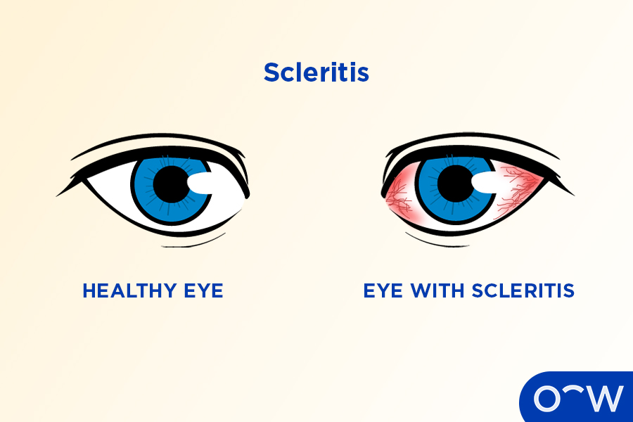
- Scleritis: Scleritis is a type of scleral disease that causes the sclera to become swollen, painful and red. Scleritis is often associated with an autoimmune disease, according to the American Academy of Ophthalmology.
- Blue sclera: As the name suggests, blue sclera occurs when the sclera is tinted a blue colour and may be caused by brittle bone disease, Marfan syndrome, iron deficiency, rheumatoid arthritis and brittle cornea syndrome.
- Eye injury: An eye injury can cause damage to the sclera resulting in blurry vision, sensitivity to light and excessive tearing. A subconjunctival haemorrhage is an eye injury caused by a scratch to the sclera, resulting in a flame-shaped bruise.
- Ocular melanosis: Ocular melanosis is a rare eye disease that results in discolouration of the sclera which may be grey, blue or brown. It most commonly only affects one eye and is often present at birth.
What are the Different Ways to Take Care of the Sclera?
The different ways to take care of your sclera include wearing protective eyewear, practising good hygiene and booking regular eye tests. The ways to take care of your sclera and their definitions are listed below.
- Wear protective eyewear: Wearing protective eyewear such as safety goggles or glasses can help protect the sclera from damage such as a scratch or an object penetrating the surface of the eye.
- Practise good hygiene: It is important to practise good hygiene such as washing your hands when touching your eyes and when putting in contacts. This can prevent bacteria and other materials from entering and irritating the eye.
- Book regular eye tests: Regular eye tests are important for the health of your sclera and overall eye health. An optometrist can check your eye health and vision for any signs of eye disease and provide treatment.
What is the Importance of Regular Visits With an Optometrist?
Regular eye tests with an optometrist are extremely important as they allow a person to stay on top of their eye health and vision. Eye tests allow an optometrist to monitor a person’s eyes to detect any signs of eye diseases or if they require prescription glasses to correct their vision. Regular visits with an optometrist should be prioritised. We suggest a person have an eye test at least once every two years.
How Can Oscar Wylee Help Take Care of Your Eye?
Oscar Wylee is able to help you take care of your eyes by providing in-store eye tests. We have Oscar Wylee optometrists who are knowledgeable and trusted to perform Medicare, bulk billed eye tests for valid cardholders using high-quality equipment. They can also provide a new or updated prescription if you need new glasses for vision correction. With same-day prescriptions, you can browse for a new pair of glasses on the same day.
Does Using Eye Drops Help the Sclera?
Eye drops can be used to manage irritation or redness on the eye's surface that affects the sclera, turning the white of the eye to red. These eye drops are called artificial tears, or lubricating eye drops and can be found at a chemist or pharmacy.
What is the Difference Between the Sclera and the Conjunctiva?
The sclera and the conjunctiva are both vital parts of the eye’s anatomy but have different functions. The sclera maintains the shape of the eyeball whereas the conjunctiva secretes mucus and tears to lubricate the eyes. The sclera is the white part of the eye and the conjunctiva is the membrane that covers the eyelids and the surface of the eye.
Read Sclera: Anatomy, Function, and Related Eye Problems in other Oscar Wylee regions and their languages.
Australia