 1300 164 990
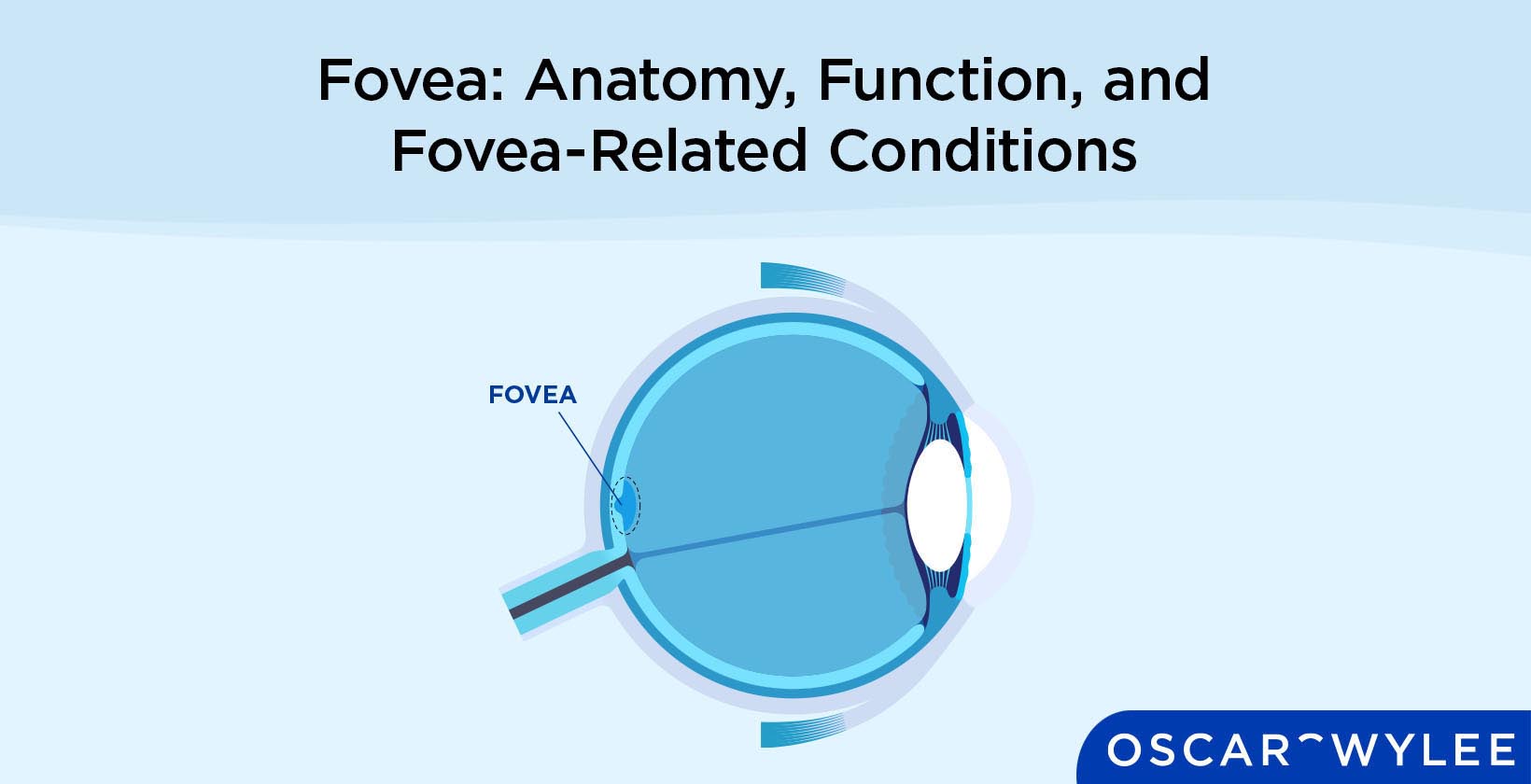

Fovea: Anatomy, Function, and Fovea-Related Conditions
Published on February 23rd, 2024
The fovea is the part of the eye’s anatomy that is responsible for a person’s highest visual acuity. The fovea, also known as the fovea centralis, is located in the macula and contains three types of cones which are long-wavelength sensitive cones, medium-wavelength sensitive cones and short-wavelength sensitive cones. Eye conditions that affect the fovea include age-related macular degeneration, diabetic retinopathy and macular telangiectasia. Keep reading to learn more about the function of the fovea centralis and its structure.
What is the Fovea?
The fovea, also known as the fovea centralis, is a small pit or depression located in the macula that provides a person’s sharpest vision or visual acuity. The fovea is located in the centre of the macula and contains three types of cones that absorb light as it enters the eye which allows them to create the sharpest possible image, according to All About Vision.
Are the Fovea and the Macula the Same Thing?
No, the fovea and macula are not the same thing, however, the fovea is located inside the macula which is inside the retina. The fovea provides the highest visual acuity whereas the macula is responsible for central vision and most of a person’s colour vision.
What is the Structure of the Fovea?
The structure of the fovea consists of three spectral types of cones, according to the National Library of Medicine. These are red or long-wavelength sensitive cones, green or medium wavelength cones and blue or short wavelength cones. The fovea is 0.35 mm in diameter and the macula is approximately 5.5 mm in diameter.

Why Does the Fovea Centralis Look Like a Pit?
The fovea centralis is a pit that is formed as the inner retinal tissue, which includes the vasculature, is moved to one side, leaving a clearer optical zone in the central foveola, according to an article published in the National Library of Medicine titled, Visual Insignificance of the Foveal Pit.
Where is the Fovea Located in Eye Anatomy?
The fovea is located in the centre of the macula lutea, which is in the centre of the posterior portion of the retina, according to an article published in the National Library of Medicine titled, Anatomy, Head and Neck, Eye Fovea. The macula is part of the retina, located in its centre at the back of the eye, therefore the answer to, is the fovea in the retina, is yes.
What Kind of Cones Are Present in the Fovea?
The cones that are present in the fovea are long-wavelength sensitive cones (L-cones), medium-wavelength sensitive cones (M-cones) and short-wavelength sensitive cones (S-cones). These cones are very compact and are thinner and more rod-like in appearance than cones elsewhere in the eye.
What is the Function of the Fovea Centralis in the Eye?
The function of the fovea contralis in the eye is to provide maximum visual acuity, meaning it allows a person to see an object with sharp vision, according to the Vision Centre Organisation. To experience this sharp vision, the eye must focus the image on the fovea centralis. Therefore, the fovea centralis function is to provide a person’s sharpest vision.
How Does the Fovea Help the Human Eye See?
The fovea helps the human eye see by providing the sharpness of a person’s vision. The fovea's role in vision works to further focus light that passes through the retina. It provides sharp vision and has a high density of cone photoreceptors that allow you to focus on details. This is called foveal vision.
Is the Fovea Centralis our Blind Spot?
No, the fovea centralis is not the eye’s blind spot, rather it provides a person’s sharpest visual acuity. According to the Vision Centre Organisation, a blind spot is where the optic nerve enters the back of the eye at the optic disc.
What are Fovea-Related Eye Problems?
Many eye problems affect the fovea including age-related macular degeneration, retinoblastoma, diabetic retinopathy, retinal detachment, macular edema, macular pucker, retinal vein occlusion, macular hole, Stargardt’s disease, macular telangiectasia and cytomegalovirus retinitis. These eye conditions and their definitions are listed below.
- Age-related macular degeneration: Both wet and dry forms of age-related macular degeneration affect the fovea. AMD is a common eye problem that causes a loss of central vision due to damage to the macula.
- Retinoblastoma: Retinoblastoma is a rare type of eye cancer that begins in the retina and commonly affects young children. Symptoms of retinoblastoma include poor vision, red eyes and eyes that appear to be looking in different directions.
- Diabetic retinopathy: Diabetic retinopathy is a complication of diabetes that can affect the fovea. This eye condition occurs when the small blood vessels in the back of the eye are damaged.
- Retinal detachment: A retinal detachment is a very serious eye condition that can affect the fovea, characterised by the retina pulling away from the layer of blood vessels that provide it with nutrients and oxygen, according to the Mayo Clinic.
- Macular edema: A macular edema occurs when blood vessels in the eye leak into the macula, causing it to swell and leading to blurry vision. Macular edema can be caused by other macula-related eye problems such as age-related macular degeneration and retinitis pigmentosa.
- Macular pucker: A macular pucker is an eye condition that occurs when bulges, wrinkles or creases form on the macula, affecting a person’s central vision. A macula pucker is commonly caused by age-related changes in the eye.
- Retinal vein occlusion: Retinal vein occlusion is a blockage of a specific vein that carries the blood away from the retina. The two types of retinal vein occlusion are central retinal vein occlusion and brand retinal vein occlusion.
- Macular hole: A macular hole is a rare condition in which there is a small break in the macula that causes blurry central vision. A macular hole is often age-related and can be treated with surgery called a vitrectomy.
- Stargardt's disease: Stargardt’s disease occurs when fatty material builds up on the macula. This is a rare genetic eye condition that may cause sensitivity to light, grey or black spots in the centre of your vision and colour blindness.
- Macular telangiectasia: Macular telangiectasia, or MacTel, is an eye disease that occurs when there are issues with the blood vessels around the fovea. The two types of MacTel are Type 1 MacTel and Type 2 MacTel, each of which affects the blood vessels differently.
- Cytomegalovirus retinitis: Cytomegalovirus retinitis is a viral eye infection of the retina, according to the American Academy of Ophthalmology. Symptoms of CMV retinitis include eye floaters and loss of peripheral vision, often leading to decreased vision.
How To Take Care of Your Fovea Centralis?
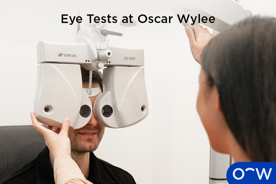
The most important way to take care of the fovea centralis is to undergo regular eye tests. Eye tests allow an optometrist to assess your vision and eye health as well as monitor your eye for any changes so they can detect eye conditions early.
What is the Importance of an Optometrist in Diagnosing Fovea-Related Eye Conditions?
An optometrist is very important in diagnosing fovea-related eye conditions and maintaining eye health. An optometrist can provide a diagnosis for age-related macular degeneration, diabetic retinopathy and retinal detachment. The role of an optometrist also includes referring patients to an ophthalmologist if these conditions require more advanced treatment.
How Important is a Regular Eye Test for the Fovea?
Regular eye tests are very important for maintaining the health of your eye including the fovea. Regular eye tests allow your optometrist to monitor any changes in your vision or eye that could indicate signs of an eye condition that may affect the fovea such as age-related macular degeneration or diabetic retinopathy.
How can Oscar Wylee Help Take Care of Your Eyes?
Oscar Wylee can help you take care of your eyes by providing comprehensive, in-store eye tests, performed by skilled and friendly optometrists. If you need glasses for vision correction, our retail team can help you find the frames and lenses that are right for your lifestyle and prescription needs. Visit an Oscar Wylee store near you to give your eyes the care they deserve.
Can You Still See Without a Fovea Centralis?
Yes, you can still see without a fovea centralis, although you will not have sharp vision, therefore, a person will have poor visual acuity, according to the Vision Centre Organisation. Your vision may also be diminished if the fovea centralis is damaged.
Does Wearing Glasses Help Protect the Fovea?
No, wearing glasses will not specifically help protect the fovea. However, wearing glasses or sunglasses in general can prevent dirt or debris from entering the eye and potentially damaging the eye. The main reason for wearing glasses is to correct a person’s vision so they can see clearly and comfortably, not to protect the fovea.
Can the Fovea Regenerate?
According to an article published in the National Library of Medicine titled, Different modes of foveal regeneration after closure of full-thickness macular holes by (re)vitrectomy and autologous platelet concentrate, the fovea may regenerate after specific surgery and injections.